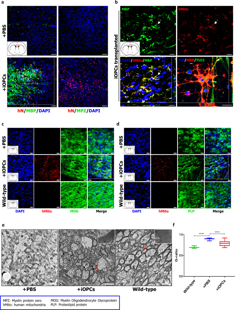
Fig. 5. Analysis of remyelination upon transplantation of iOPCs in shiverer mice.
a Shiverer mice were implanted in the corpus callosum with O4+ iOPCs or treated with PBS. The transplantation of iOPCs into the corpus callosum resulted in their differentiation into MBP-positive oligodendrocytes. Transplanted iOPCs were identified using a human-specific nuclear antibody. Scale bars, 100 µm. Higher-magnification fluorescence images of MBP- (b), MOG- (c), and PLP- (d) positive engrafted cells. Engrafted iOPCs were identified using a human-specific anti-mitochondrial antibody. Scale bars, 20 µm. e Representative electron microscope images of PBS-treated brains of shiverer mice, iOPC- engrafted brains of shiverer mice, and untreated brains of wild-type mice. While the engrafted corpus callosum in iOPC-transplanted and wild-type brains show compact myelin with major dense lines, the PBS-treated brains contained thin myelin sheaths. The samples of the corpus callosum were obtained 14 weeks after engraftment. The arrows denote the original position of magnified picture. Scale bars, 2 µm. f The comparison of the g-ratio in the corpus callosum among the PBS-treated brains of shiverer mice, iOPC- engrafted brains of shiverer mice, and untreated brains of wild-type mice. The graph represents the g-ratio obtained from axons with a diameter >1 µm and was calculated from three mice per group (n = 100). *, statistically significant difference vs. PBS-treated group. Significant differences were analyzed by one-way ANOVA (iOPCs, ****P < 0.0001; Wild-type, ****P < 0.0001).

