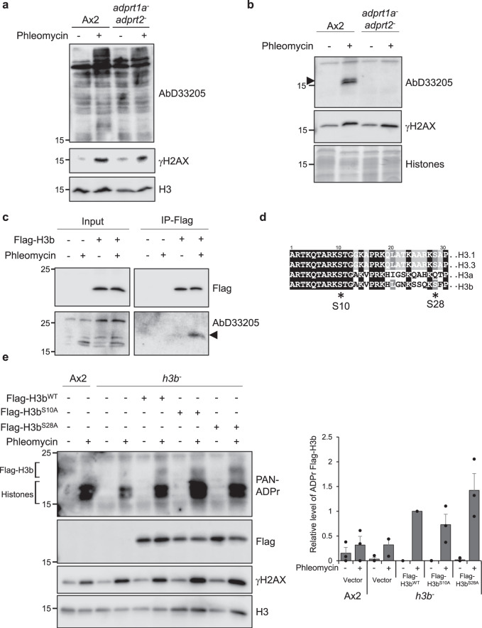
Fig. 2. H3b is ADP ribosylated on serine in response to DNA damage.
a, b Ax2 or adprt1a−adprt2− cells were treated with phleomycin and whole cell (a) or acid (b) extracts western blotted with antibodies as indicated. Molecular weight markers are indicated in kDa. Representative pictures of three biological repeats are presented. c h3b− cells containing empty vector, or expressing Flag-H3bwt, were left untreated or exposed to phleomycin. Following preparation of denatured chromatin, Flag immunoprecipitation was performed. Input extracts or immunoprecipitates were subjected to Western blotting using the indicated antibodies. Molecular weight markers are indicated in kDa. Representative pictures of at least three independent experiments. d Sequence comparison of human histone H3.1 and H3.3 with the Dictyostelium H3 variants H3a and H3b. S10 and S28, the main ADP-ribosylation targets in vertebrates are indicated. e Ax2 or h3b− cells containing empty vector, or expressing Flag-H3bWT, Flag-H3bS10A or Flag-H3bS28A were left untreated or exposed to phleomycin. Following preparation of acid extracts, western blotting was performed using the indicated antibodies. ADP-ribosylated endogenous histones and Flag-H3b are highlighted. Enrichment of Flag-H3b in chromatin fractions was quantified (right panel; n = 3; individual data points are shown and error bars represent the SEM). Molecular weight markers are indicated in kDa. Source data are provided in the Source Data file.

