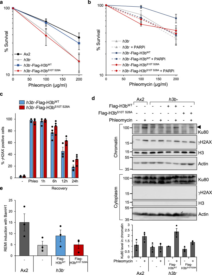
Fig. 4. H3b ADP-ribosylation is required for DSB repair.
a Ax2 cells, or h3b− cells expressing Flag-H3bWT or Flag-H3bS10T S28A were exposed to phleomycin at the indicated concentrations and cell viability assessed by clonogenic survival assays (data represent seven biological repeats; error bars represent the SEM). b h3b− cells alone, or expressing Flag-H3bWT or Flag-H3bS10T S28A were exposed to phleomycin in the absence or presence of PARP inhibitors (olaparib; PARPi) as indicated and cell viability assessed by clonogenic survival assays (data represent three biological repeats; error bars represent the SEM). c h3b− cells expressing Flag-H3bWT or Flag-H3bS10T S28A were left untreated (−) or exposed to phleomycin for 1 h (Phleo). Following removal of phleomycin, recovery of cells was analysed at the time points indicated. DNA damage was assessed by scoring γH2AX nuclei with >5 foci (n = 3; individual data points are shown and error bars represent the SEM). d Ax2, h3b− cells, or h3b− cells expressing Flag-H3bWT or Flag-H3bS10T S28A were left untreated or exposed to phleomycin as indicated. Cytoplasmic and chromatin fractions were prepared from cells and western blotting performed using the indicated antibodies (left panel). Molecular weight markers are indicated in kDa. Enrichment of Ku80 in chromatin fractions was quantified (lower panel; n = 3; individual data points are shown and error bars represent the SEM). e Restriction-Enzyme Mediated Integration REMI of plasmid DNA was evaluated in Ax2, h3b− cells, or h3b− cells expressing Flag-H3bWT or Flag-H3bS10T S28A (n = 3; individual data points are shown and error bars represent the SEM). Source data are provided in the Source Data file.

