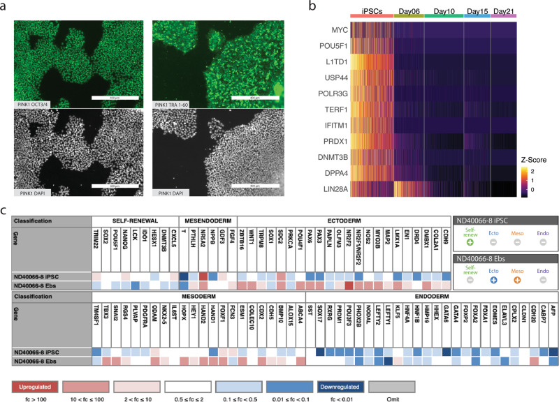
Fig. 2. Classification of iPSC status.
a Immunocytochemistry (ICC). Staining for the iPSC markers POU5F1 (more commonly known as OCT3/4) and TRA-1-60 of iPSC colonies, prior to differentiation. DAPI was used to stain cell nuclei as a reference. b Expression of genes known to indicate iPSC status (MYC and POU5F1) and of genes identified by a differential expression analysis between iPSCs and differentiating cells (also see Supplementary Fig. 12). Colors correlate to normalized counts (z-score, centered, and scaled) of the indicated genes. TDGF-1 is expressed in iPS cells of high stemness;41 L1TD1, USP44, POLR3G, and TERF1 are essential for the maintenance of pluripotency in human stem cells;47–50 IFITM1, PRDX1, DNMT3B, DPPA4, and LIN28A and are associated with stemness51–53,137,138. c Results of Scorecard analysis of iPSCs and embryonic bodies (EBs)44,45. iPSCs are expected to show high expression of self-renewal genes and low expression of mesoderm, ectoderm, and endoderm markers. EBs are cells at an early stage of spontaneous differentiation. Scorecard analysis of EBs determines the iPSC line’s potential to differentiate into the three germ layers, hence, EBs are expected to express few or no self-renewal genes and to show expression of some mesoderm, ectoderm and endoderm markers: Ecto±, Meso±, Endo±.

