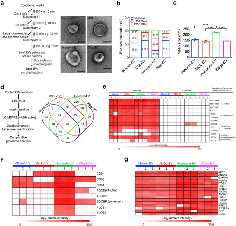
FIGURE 2.

Isolation and characterization of extracellular vesicles from hiPSC‐derived brain cells. (a) Workflow for the EV isolation and transmission electron microscopy (TEM) images of isolated iNeuron‐, iMGL‐, iAstrocyte‐, and iOligo‐EVs. Scale bar: 100 nm. (b) Size distribution of the isolated EVs from four cell types determined using a nanoparticle tracking analysis (NTA, n = 3 for each cell type). (c) Comparison of the EV mean size among the four cell types by NTA (n = 3 for each cell type). iAstrocyte showed the significant increase in diameter of the EV size when compared to the other three cell types. Data are presented as the mean ± SEM. **** p < 0.0001. (d) Workflow for the label‐free proteomics and Venn diagram showing the number of EV proteins differentially identified in iNeuron, iMGL, iAstrocyte, and iOligo. (e) Heatmap illustrating the expression of non‐EV protein markers present in different brain cell‐derived EVs and their cellular origins based on the protein intensity determined using mass spectrometry. Four types of non‐EV protein markers, including proteins from the nucleus, mitochondria, secretory pathway, and others, were selected as indicated in the MISEV2018 guideline (Théry et al., 2018). (f) Heatmap illustrating the enrichment of conventional exosome protein markers present in different brain cell‐derived EVs based on their protein intensity from mass spectrometry. (g) Sixteen newly defined pan‐EV marker candidates from different cell types based on their protein intensity, determined using mass spectrometry. Proteins represented in at least two of the three replicates within each cell type were selected. The white cells indicate no presence of the protein in the specified cell type. hiPSC, human induced pluripotent stem cells; EV, extracellular vesicle; TEM, transmission electron microscopy; iNeuron, hiPSC‐derived excitatory neuron; iMGL, hiPSC‐derived microglia‐like cell; iAstrocyte, hiPSC‐derived astrocyte; iOligo, hiPSC‐derived oligodendrocyte‐like cells; NTA, nanoparticle tracking analysis; SEM, standard error of the mean; MISEV2018, Minimal information for studies of extracellular vesicles 2018
