Figure 4.
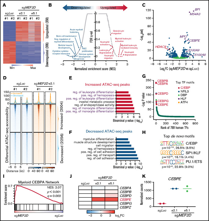
MEF2D represses genes involved in the CEBPE-centered myeloid differentiation. (A) Transcriptomic analysis of differentially expressed genes (fold change >1.5 and adjusted p-value <.05) between MEF2D-knockout and control MOLM-13 cells. (B) Gene sets enrichment analysis (GSEA) revealing top enriched gene signatures in upregulated and downregulated genes in MEF2D-knockout MOLM-13 cells. (C) Volcano plot showing fold change and adjusted p-value of gene expression between MEF2D-knockout and control MOLM-13 cells. Differentially expressed genes are marked in blue. (D) Heatmap displaying the ATAC-seq read densities in peaks with increased or decreased chromatin accessibility in MEF2D-knockout MOLM-13 cells, compared with sgLuc control. (E-F) Genomic Regions Enrichment of Annotations Tool annotation of ATAC-seq peaks with increased (E) or decreased (F) chromatin accessibility as identified in panel D. (G) Motifs enriched in increased ATAC-seq peaks against 769 human transcription factors. Top motifs were highlighted and labeled. (H) Top de novo motifs identified from increased ATAC-seq peaks. (I) GSEA revealing an enrichment of C/EBP network genes in MEF2D-knockout cells. (J) Heat map showing fold change of expression of C/EBP family genes upon MEF2D knockout. FC, fold change. (K) Normalized RNA-seq read counts of CEBPE in MOLM-13 cells harboring sgRNAs against Luciferase gene or MEF2D.

