Figure 6.
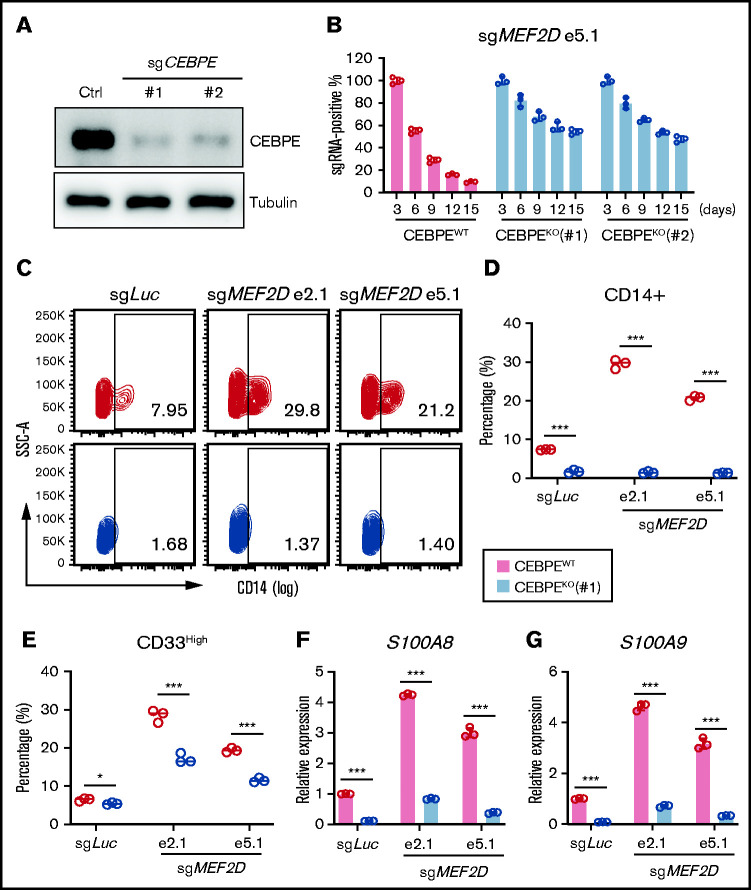
CEBPE depletion can partially rescue phenotypes of MEF2D loss. (A) Western blot analysis demonstrating efficient deletion of CEBPE after stable knockout of CEBPE using 2 independent guide RNAs, relative to empty vector control. (B) Competitive proliferation assay demonstrating CEBPE knockout partially rescues MEF2D loss-induced proliferation defect. MEF2D knockout was performed in CEBPE wildtype (WT) and 2 independent knockout (KO) lines by using sgRNA e5.1 linked with an mCherry reporter. The mCherry positive percentage was monitored every 3 days post infection and normalized to the day 3 measurement. (C) Flow cytometry analysis of myeloid differentiation marker CD14 in CEBPE wildtype and knockout MOLM-13 cells 6 days post MEF2D knockout, relative to transduction of sgLuc non-targeting control. (D-E) Flow cytometry analysis showing frequency of population with high myeloid differentiation markers CD14 (D) and CD33 (E) in CEBPE wildtype and knockout MOLM-13 cells 6 days post MEF2D knockout, relative to transduction of sgLuc non-targeting control. (F-G) RT-qPCR analysis of CEBPE target genes S100A8 (F) and S100A9 (G) in CEBPE wildtype and knockout MOLM-13 cells 6 days post MEF2D knockout, relative to transduction of sgLuc non-targeting control. *P < .05; ***P < .001.

