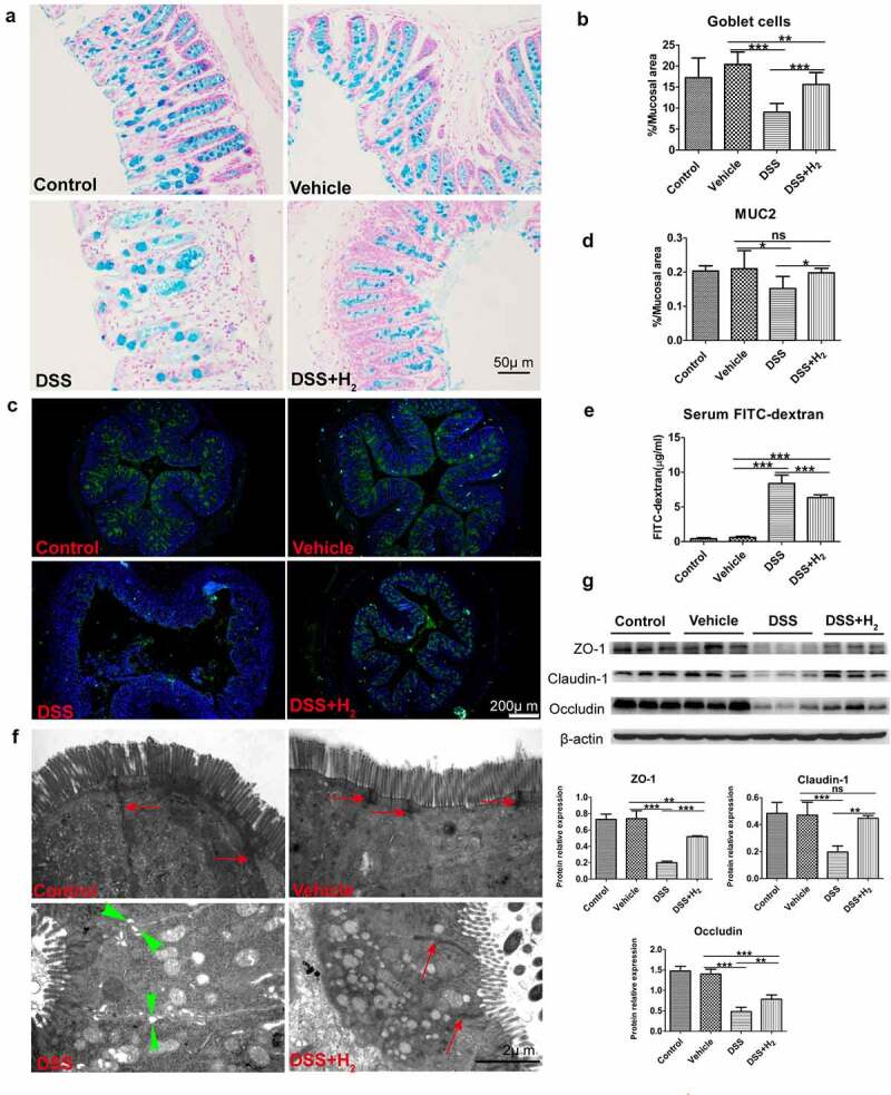
Figure 9.

HS administration reinforces mucous layer architecture and integrity of the epithelial barrier during colitis. (a-b) Alcian blue staining for goblet cells in colon tissue and quantification of staining intensities (n = 5). (Scale bars: 50 μm). (c-d) Immunofluorescence staining for α-MUC2 (green) and nuclei (blue) and quantification of staining intensities (n = 5). (Scale bars: 200 μm). (e) In vivo epithelial barrier permeability (n = 5). (f) Representative TEM micrographs showing the ultrastructure of colonocytes(Scale bars: 2 μm). Red arrows with shafts delineate tight junction domains and opposing green triangular arrowheads point to intercellular space between two neighbored cells (g) Western blot was used to respectively determine intestinal interepithelial tight junction proteins including Occludin, Claudin-1 and ZO-1 with β-actin as reference in colon tissue (n = 3). All data show mean ± SD, are representative of two to four independent experiments, and include statistical significance calculated by one-way ANOVA with LSD multiple comparison post hoc test. ns, no significant; * P < .05; **P < .01; ***P < .001.
