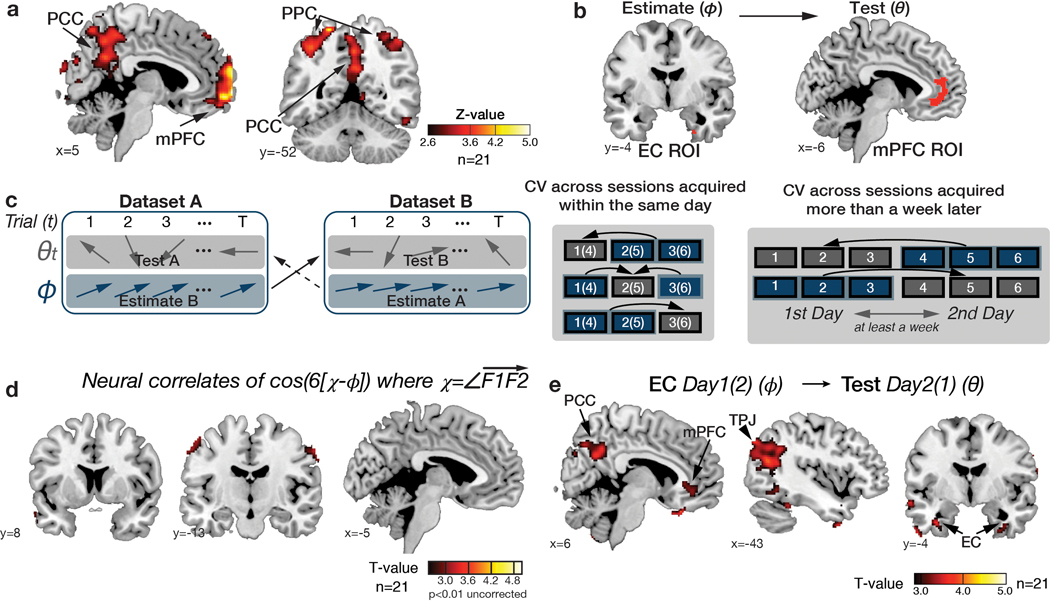
Extended Data Fig. 3. The analysis procedure to examine the effects of grid-like codes.
a. Identifying brain regions sensitive to hexagonal symmetry across the whole brain without aligning to the entorhinal grid orientation, φ. To identify hexagonally symmetric signals, we adopted previously developed methods from a previous study 14. We used a Z-transformed F-statistic to examine the BOLD activity modulated by any linear combination of sin(6θ) and cos(6θ). Hexagonal modulation was found in brain regions including in medial prefrontal cortex (mPFC; peak MNI coordinate, [x,y,z]=[2,66,−4], z=5.58), posterior cingulate cortex/precuneus (PCC; [2,−50, 36], z=3.19), bilateral posterior parietal cortex (PPC; [36,−46, 62], z=4.27 for right; [−38,−50, 50], z=3.81 for left), left lateral orbitofrontal cortex (OFC; [−42,44,−8], z=3.79), and retrosplenial cortex (RSC; [x,y,z] = [2,−54,28], z = 3.45) at a threshold pTFCE<0.05 (whole brain TFCE correction), as well as the right entorhinal cortex (EC; [26,−10, −40], z=2.80) at a threshold, pTFCE<0.05 (corrected within a priori anatomically defined EC region of interest (ROI) 62,63). For visualization purposes, the maps are thresholded at z>2.6 (p<0.005 uncorrected). b. We did not use the results of F-test for statistical inference but to functionally define ROIs in EC and mPFC for future tests. The ROIs were defined within the anatomically defined masks in the EC 62,63 and mPFC 72 by including effects at a threshold of z>2.3, which corresponded to p<0.01. Using these independently defined ROIs, we then tested if the grid orientation estimated in the ROI was consistent across separate fMRI sessions in an unbiased way. It is important to note that the ROIs were defined from results of a statistically independent analysis, which was not dependent on the grid orientation. c. Illustrations of the cross-validation (CV) procedure. By splitting a dataset for estimating the grid orientation, φ, from another dataset to test for hexagonal modulation for inferred trajectories, φ, we could test for brain regions where activity was modulated by cos(6[θ−φ]) in alignment with the grid orientation estimated from the independent dataset for each participant. The CV was possible because the grid orientation, φ, is thought to be relatively stable but different across participants, whereas the direction of inferred trajectories varies only according to the position of F0, F1, and F2 (left panel). We performed a CV procedure both from fMRI sessions acquired within the same day (middle panel), and also from fMRI sessions acquired more than a week apart (right panel). d. We estimated the angle of the F1F2 vector (χ) and inputted the cos(6[χ − φ]) at the time of F2 presentation as an additional parametric regressor into GLM2. We did not find any brain areas encoding the angles of F1F2 vectors, except at a reduced threshold, in the bilateral somatosensory cortex ([x,y,z] = [−52,−14, 56], t=3.33 and [58,−8,50], t=3.17, p<0.005 uncorrected). e. Consistency of grid orientation in EC across sessions acquired more than a week apart. Concretely, in alignment with the EC grid orientation estimated from sessions acquired on a different day, we found hexadirectional modulation in a network of brain regions, including the mPFC ([−2, 36, −8], t=5.04), PCC ([6,− 58, 30], t=3.68), and TPJ ([−36, −64, 22], t=4.06) at our whole brain corrected threshold pTFCE<0.05, as well as in EC ([36, −10, 38], t=4,21) at pTFCE<0.05, small-volume-corrected in our a priori EC ROI (Supplementary Table 5b).

