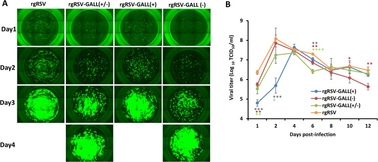
Fig 6. RSV replication in HBE cultures.
(A) Spreading of m6A-deficient rgRSVs in HBE culture. HBE cultures were infected by 400 TCID50 of each rgRSV mutant. At the indicated time, virus spread was monitored by fluorescence microscopy. Representative micrographs at each time point are shown. (B) Virus release from m6A-deficient rgRSV-infected HBE culture. HBE cultures were infected by 400 TCID50 of each rgRSV. After virus inoculation, supernatants were collected on day 1 and every 2 days until day 12 post-inoculation. Infectious virus in supernatants was determined by TCID50 assay. Viral titers are the geometric mean titer (GMT) of three independent experiments ± standard deviation. Data were analyzed using Student’s t-test and *P < 0.05; **P < 0.01; ***P < 0.001; ****P < 0.0001.

