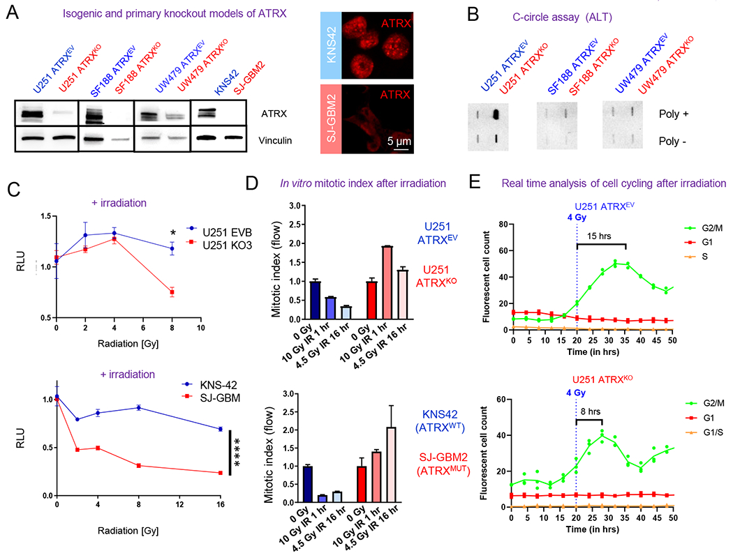
Figure 1. ATRX-deficient human glioma cells demonstrate inappropriate cell cycling after treatment with irradiation.

(A) Western blot of U251, SF188 and UW479 isogenic ARTXKO cells illustrating ATRX reduction. Immunoblots of KNS42 and SJ-GBM2 are also included (right two columns) illustrating ATRX reduction in ATRX-mutant SJ-GBM2 cells and immunocytochemistry images (right image) demonstrating their punctate nuclear staining of ATRX only in ATRX-wildtype KNS42 cells. (B) C-circle assay results demonstrating DNA from each sample in the presence (+) or absence (−) of Φ29 polymerase; positive staining demonstrates ALT in U251-ATRXKO cells only. (C) In vitro data showing proliferation of U251 ATRXKO cells and its isogenic control, as well as KNS42 (ATRX wildtype) and SJ-GBM2 (ATRX mutant), after exposure to increasing doses of irradiation (IR). (D) In vitro mitotic index assay at 1 hour after 10 Gy IR and 16 hours after 4.5 Gy IR demonstrates that U251 (ATRXKO) and SJ-GBM2 (ATRX mutant) cells display increased proliferation following treatment with IR. All values normalized to respective 0 Gy value. 0 Gy is normalized to 1.0. (E) Real-time analysis of cell cycle transitions using the Incucyte imaging system and the FastFUCCI reporter plasmid shows the U251 ATRXKO cells return to active cycling 2X faster after 4 Gy IR. [Mean ± SEM for triplicate experiments are shown. *P≤0.05, **P≤ 0.01, ***P≤0.001, and ****P≤0.0001 using 2-Way ANOVA.] For additional data, see also Figure S1.
