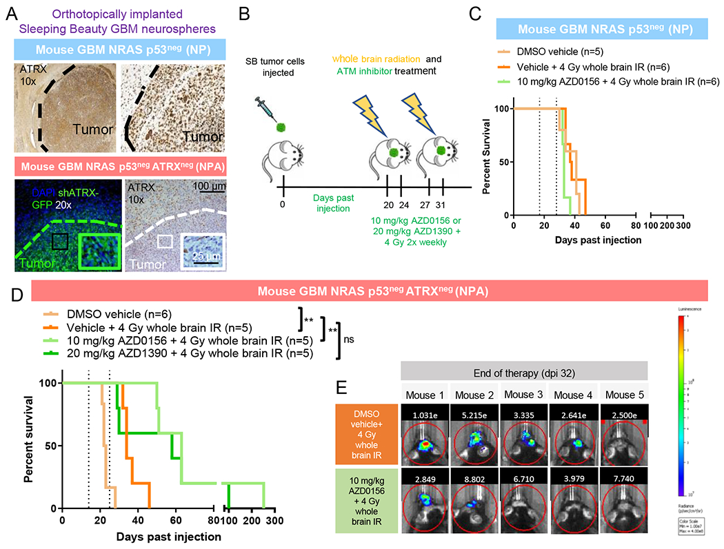
Figure 7. Treatment of implanted ATRX-deficient mouse GBM cells with ATM inhibitors.

(A) Immunofluorescence and IHC staining of tumors of mice implanted with mouse GBM neurospheres with (NPA) and without (NP) ATRX knockdown (GFP stains for shATRX plasmid). Tumor shows robust ATRX loss which is not seen in surrounding normal cortex. Boundary between tumor and non-tumor tissue is denoted by dotted line. (B) Schematic of treatment with whole brain IR and ATM inhibitor for mice implanted with tumor cells. (C) Kaplan-Meier survival curves of C57BL/6 mice bearing SB implanted NP tumors with no response to irradiation +/− ATM inhibition. (D) Kaplan-Meier survival curves of C57BL/6 mice bearing SB implanted NPA tumors show significant radio-sensitization with AZD0156. **P≤ 0.01 using Log-rank (Mantel-Cox) test. (E) Tumor bioluminescence for tumor-bearing NPA mice treated with 4 Gy whole brain irradiation with and without AZD0156. For additional data, see also Figure S7.
