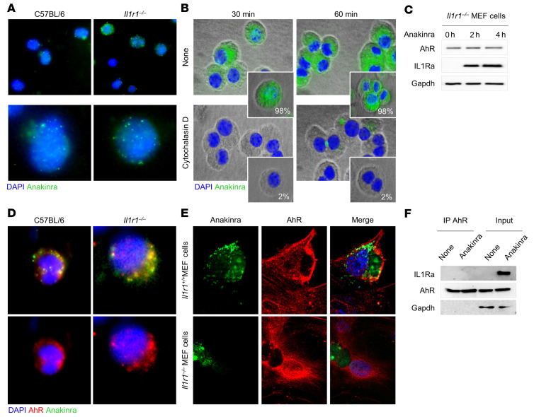
Figure 4. Anakinra colocalizes with AhR in Il1r1–/– cells.
(A) Immunofluorescence imaging of cellular localization of FITC-anakinra in alveolar macrophages, isolated from naive C57BL/6 or Il1r1–/– mice, primed with 100 ng/mL LPS for 2 hours at 37°C, and exposed to 10 μg/mL FITC-anakinra for 60 minutes. (B) Immunofluorescence analysis of FITC-anakinra in Il1r1–/– cells exposed to FITC-anakinra at 30 and 60 minutes in the presence of 5 μM cytochalasin D. Insets, percentage positive cells. (C) Il1r1–/– MEF cells were treated with 10 μg/mL anakinra for 0, 2, and 4 hours and assessed for AhR and IL1Ra expression by immunoblotting with specific antibodies. (D) Immunofluorescence imaging of alveolar macrophages from naive C57BL/6 and Il1r1–/– mice primed with 100 ng/mL LPS for 2 hours at 37°C, exposed to 10 μg/mL FITC-anakinra for 60 minutes, and stained with anti-AhR antibody. (E) Immunofluorescence imaging of MEF cells stained with anti-AhR antibody and DAPI and exposed to 10 μg/mL FITC-anakinra for 60 minutes. (F) Il1r1–/– MEF cells were treated with 10 μg/mL anakinra, lysed, immunoprecipitated with anti-AhR antibody, and assessed for IL1Ra and AhR expression by immunoblotting with specific antibodies. None, untreated cells. Representative images, acquired with a fluorescent microscope (BX51), and immunoblots from 2 independent experiments are shown. DAPI was used to detect nuclei. Sections were examined using a Zeiss Axio Observer Z1.

