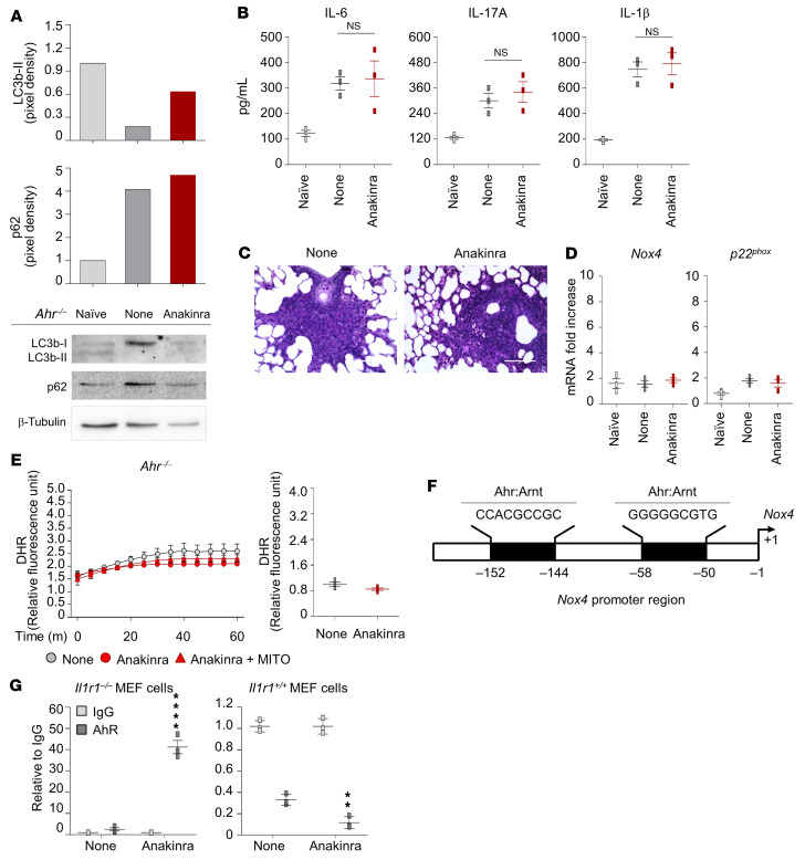
Figure 6. Anakinra promotes AhR transcriptional activity.
(A–C) AhR–/– mice were infected and treated with anakinra as in legend to Figure 1 and assessed for immunoblotting of LC3b and p62 (A), cytokine production (ELISA) in lung homogenates (B), and lung histology (PAS staining) at 7 days after infection (C). Scale bar: 200 μm. Data represent 3 independent experiments. Each in vivo experiment includes 6 to 8 mice per group, pooled before analysis. NS, not statistically significant, treated versus untreated (None) cells. One-way ANOVA, Bonferroni post hoc test. (D and E) Ex vivo purified alveolar macrophages and total lung cells from Ahr–/– mice infected and treated with anakinra were assessed for Nox4 and p22phox expression (RT-PCR) (n = 3 independent samples) (D) and H2O2 production (DHR staining) (n = 2 independent samples) (E). (F) Illustration of predicted binding sites from the ALGGEN-PROMO database and the Eukaryotic Promoter Database. (G) Il1r1–/– and Il1r1+/+ MEF cells were treated with 10 μg/mL anakinra. ChIP assay was performed with AhR antibody. IgG was used as negative control. qPCR was conducted at the promoter regions of Nox4. Data are technical replicates of 1 representative out of 2 independent experiments.

