Figure 4.
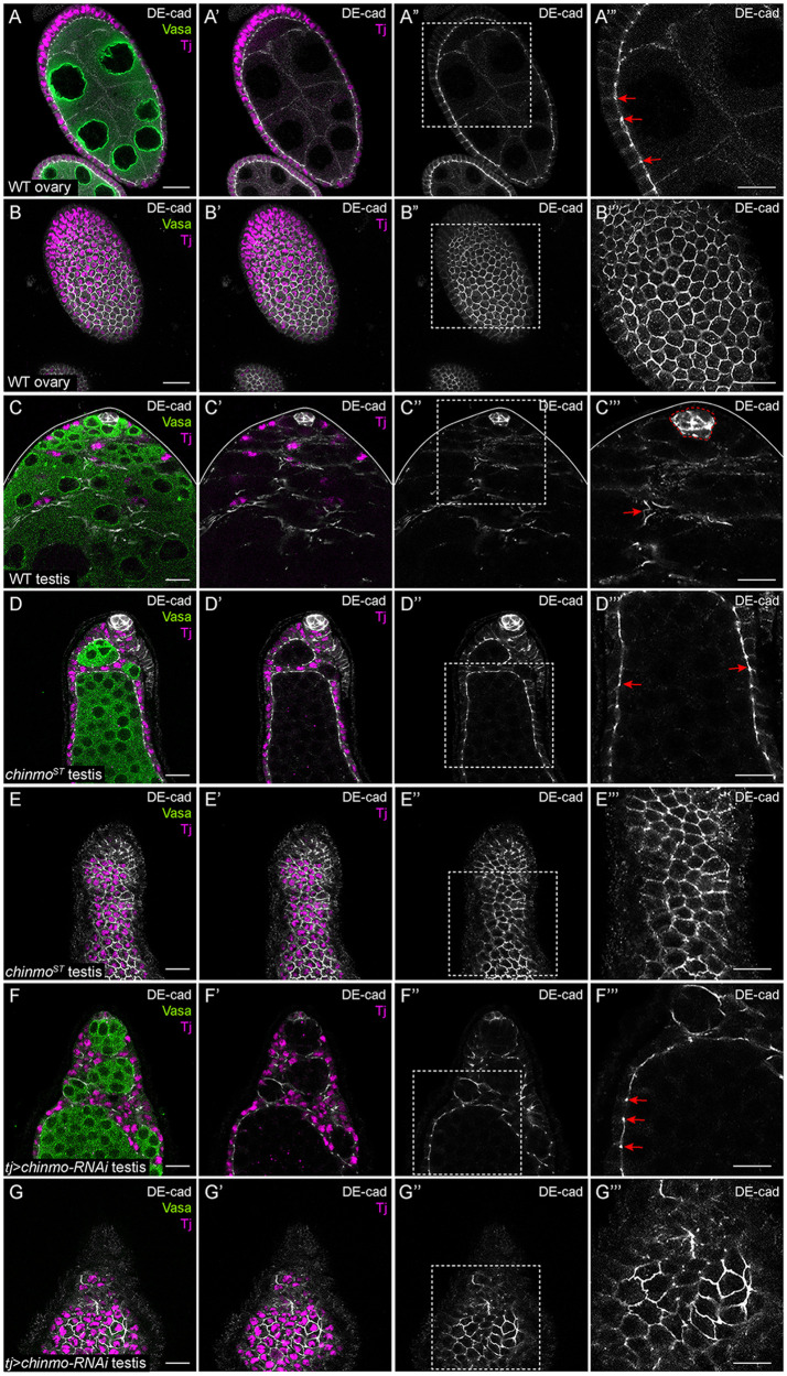
chinmo-deficient somatic cells upregulate DE-cad. (A–A’”) Middle xy-section of a WT ovary shows that DE-cad (white) is expressed at AJs (arrows in A’”) in somatic follicle cells. (B–B’”) An apical xy-section of a WT ovary shows that DE-cad (white) is apically enriched in follicle cells. (C–C”’) In a WT testis, DE-cad (white) is expressed strongly in the niche (outlined by a dashed line in C’”) and at a modest level in the somatic cells (arrow, C’”). (D–D”’) Middle xy-section of a chinmoST mutant testis shows that DE-cad (white) is ectopically expressed at AJs (arrows, D’”) in feminized somatic cyst cells. (E–E’”) An apical xy-section of a chinmoST mutant testis reveals apical enrichment of DE-cad (white) in the feminized somatic cells. (F–F’”) Middle xy-section of a tj>chinmo-RNAi testis shows that DE-cad (white) is ectopically expressed at AJs (arrows, F’”) in feminized somatic cyst cells. (G–G’”) An apical xy-section of a tj>chinmo-RNAi testis shows apical enrichment of DE-cad (white) in the feminized follicle-like cells. In (A–G), Vasa (green) marks the germline and Tj (magenta) marks cyst cells. Scale bar = 10 μm. (A’”–G”’) is a magnification of the boxed regions in (A”–G”), respectively. Time point in (A–G) is 7–8 days post-eclosion. Genotypes: (A, B) +/+; +/+; +/+ (OregonR), (C) +/Y; +/+; +/+ (OregonR), (D, E) w/Y; chinmoST/chinmoST; +/+ (F, G) w/Y; tj-Gal4/+; UAS-chinmo-RNAi/UAS-Dcr-2 (labeled “tj>chinmo-RNAi”).

