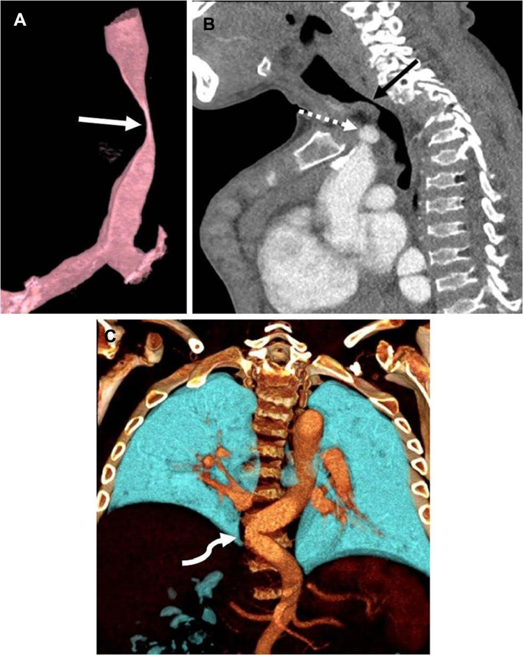
Figure 1.
Computed tomography angiogram in a 16-year-old boy with mucopolysaccharidosis type IVA. (A) Three-dimensional reconstruction of the trachea in a sagittal oblique projection shows a severe narrowing of the trachea at the thoracic inlet (arrow). (B) Sagittal image shows that the brachiocephalic artery (dashed arrow) contributes to crowding at the thoracic inlet but does not directly indent the trachea (solid arrow). (C) Three-dimensional coronal reconstruction posteriorly in the chest shows tortuosity of the descending thoracic aorta, crossing the midline (arrow). Reprinted by permission from Springer Nature, Averill LW, Kecskemethy HH, Theroux MC, et al. Tracheal narrowing in children and adults with mucopolysaccharidosis type IVA: evaluation with computed tomography angiography. Pediatr Radiol. 2021;51(7):1202–1213. Figure 1 is copyright protected and excluded from the open access licence.18

