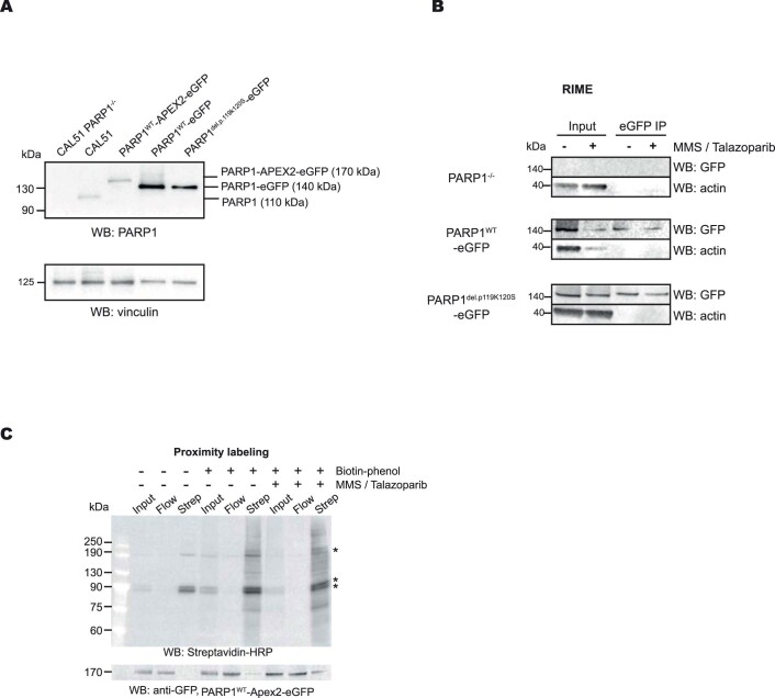
Extended Data Fig. 1. Proteomic profiling of PARP1 transgene-expressing CAL51 cells.
a. Western blot showing the expression of PARP1 transgenes, detected by an PARP1 antibody. Data shown represent 2 biological replicas. b. A Western blot analysis of the purified PARP1-associated proteins as described in the RIME experiment in Fig. 1a. Data shown represent 3 biological replicas. c. Western blot analysis of the purified biotinylated proteins isolated in the PARP1WT-Apex2-eGFP proximity labelling experiment. Immunoblotting using Streptavadin-HRP is shown in the top panel, whilst anti-GFP immunoblotting is shown in the bottom panel. Endogenously biotinylated proteins are indicated as*. Data shown represent 3 biological replicas.

