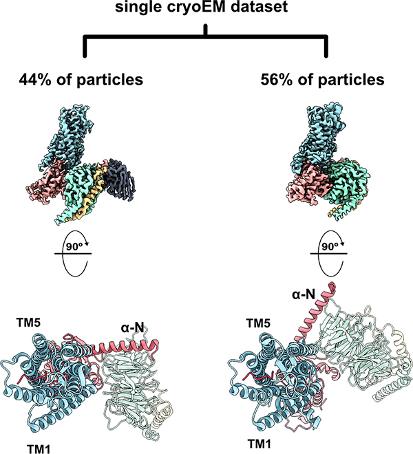
Figure 4: From a single cryoEM dataset, 3-dimensional classification of projections revealed two distinct conformers representing two distinct GPCR-G protein interaction states representing two thermodynamically comparable conformers.
In the canonical state (left, PDB ID 6OS9), the receptor engages the G protein in a prototypical fashion in which the nucleotide binding pocket is primed for GTP binding. In the non-canonical state (right, PDB ID 6OSA), the G protein heterotrimer is rotated by 45° compared to the canonical state, representing an intermediate ligand-bound receptor state along the G protein coupling pathway. TM: transmembrane helix; α-N: N-terminal alpha helix of G protein.

