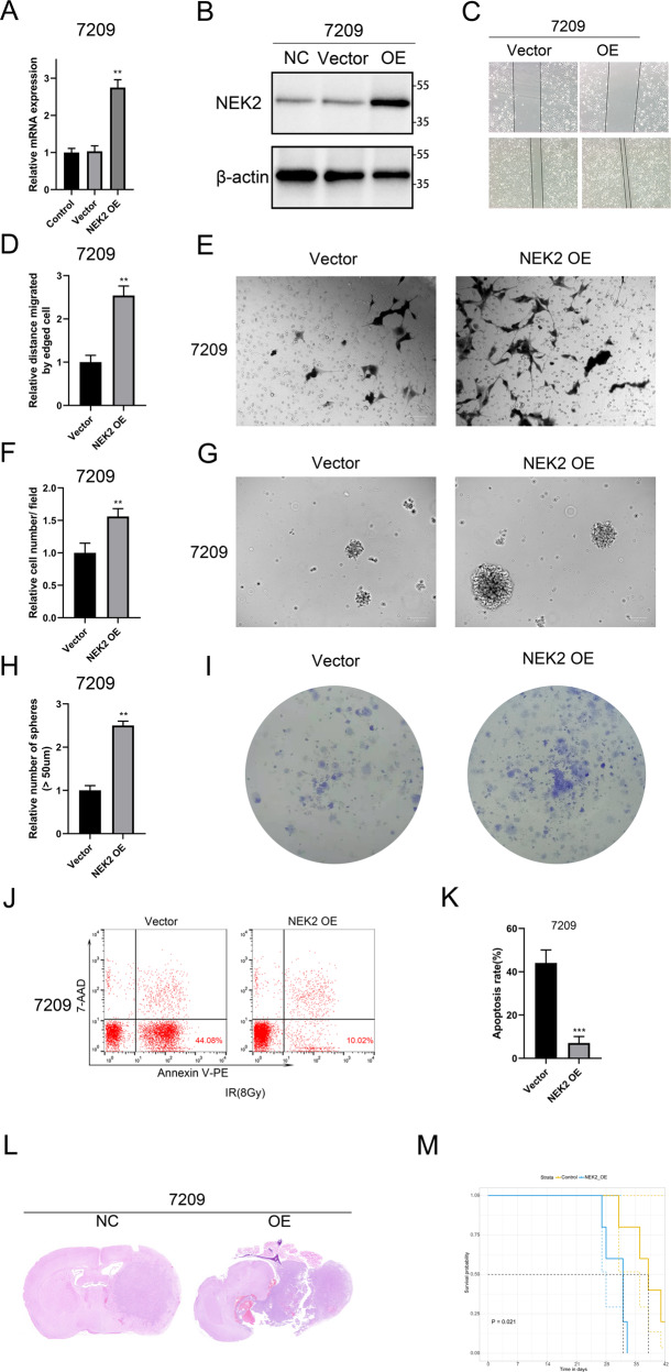
Fig. 3. NEK2 overexpression promoted GBM progression both in vitro and vivo.
A Analysis of the mRNA expression of NEK2 in pretreatment group and control group by qRT-PCR. B Western blot analysis for detecting the NEK2 protein expression in 7209 cells transduced with lentiviral NEK2, lentiviral vector and blank control. C, D Wound healing assays for exploring the effect of NEK2 overexpression on tumor malignancy in GBM cells. E, F Invasive ability of GBM cells transduced with lentiviral NEK2 and control. G, H Sphere formation assays were performed to detect the sphere formation efficiency of NEK2 overexpression cells and control cells. I Colony formation assays to verify the effect of NEK2 overexpression on the proliferation of GBM. J, K Flow cytometry analysis using Annexin V and Propidium Iodide for apoptotic ratio analysis in 1763 cells transduced with lentiviral NEK2 and control. L Representative H&E-stained images of mouse brain sections after the intracranial transplantation. Scale bars: 3 mm. M Kaplan-Meier survival curves of the xenograft mice in control group and NEK2 overexpression group (P = 0.021, with log-rank test). All data were presented as the mean ± SD of triplicate independent experiments.

