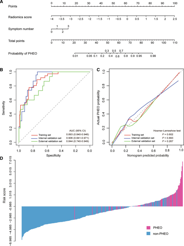
Fig. 3.
The radiomic-clinical nomogram and its performance. A The radiomic-clinical nomogram was developed to distinguish PHEOs from other adrenal lesions. B ROC curves of the radiomic-clinical nomogram in the training, internal and external validation sets. C Calibration curves of the nomogram in the training, internal and external validation sets. The calibration curve presents how well the predicted probabilities agree with the observed probabilities. The diagonal dotted line indicates the ideal prediction by the ideal model. The solid lines present the prediction value of the nomogram. A closer fit of the solid line to the diagonal dotted line demonstrates a better prediction. D The calculated risk scores for each patient within the combined training, internal and external validation datasets

