FIGURE 2.

Automated MBF Pixel Maps in a Normal Volunteer
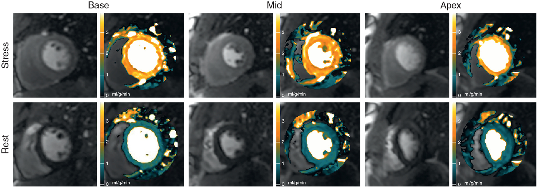
Automated stress and rest myocardial blood flow (MBF) pixel maps in a normal volunteer show coherent hyperemic MBF (orange) and rest MBF (green) on all 3 slices (Online Video 1).

Automated MBF Pixel Maps in a Normal Volunteer
Automated stress and rest myocardial blood flow (MBF) pixel maps in a normal volunteer show coherent hyperemic MBF (orange) and rest MBF (green) on all 3 slices (Online Video 1).