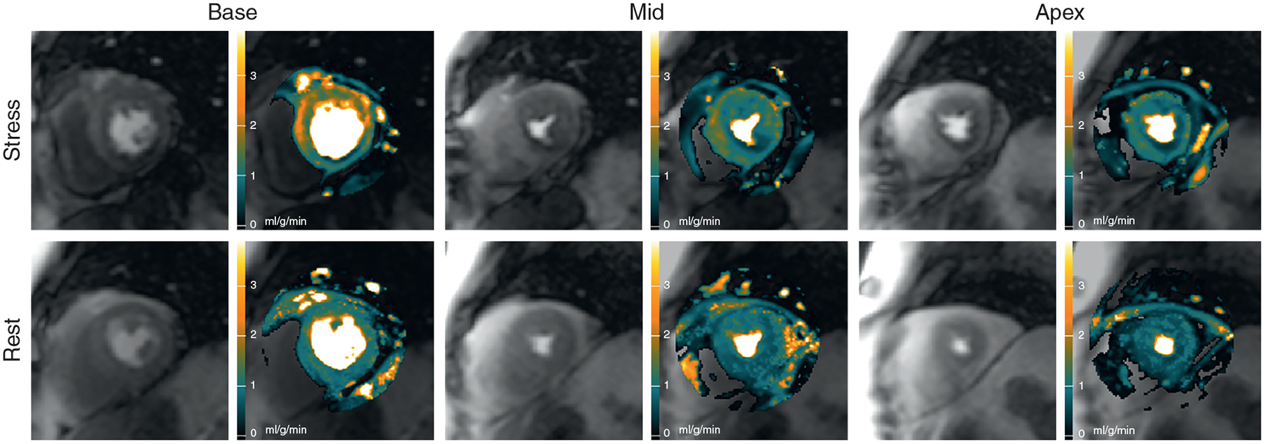
FIGURE 4.

Automated MBF Pixel Maps in Patient With Multivessel Disease
Automated MBF pixel maps in a patient with an 87% circumflex stenosis, an 84% left anterior descending stenosis, and a 65% right coronary artery stenosis by QCA show corresponding perfusion defects in the stress maps in all 3 coronary artery territories. There is some epicardial hyperemic perfusion in the basal anteroseptal and anterolateral segments (Online Video 3). MBF = myocardial blood flow; QCA = quantitative coronary angiography.
