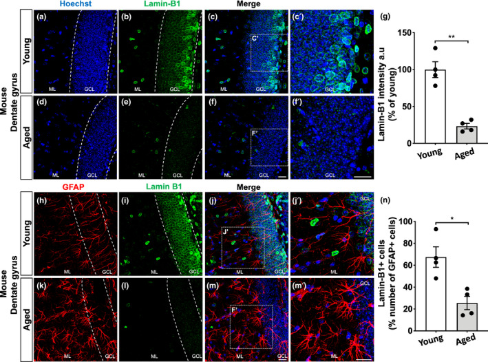
FIGURE 1.

Age‐related loss of lamin‐B1 in the mouse hippocampus. (a–g) Densitometric analysis of lamin‐B1 staining in the mouse hippocampal dentate gyrus, including the molecular layer (ML) and granular cell layer (GCL), revealed a global reduction of lamin‐B1 intensity in aged mice compared with young mice (p = 0.0033). (h–n) Decreased proportion of lamin‐B1 + cells in relation to the total number of GFAP + cells in the dentate gyrus of aged mice compared with young ones (p = 0.0126). Significance was determined using unpaired t test with Welch's correction. Error bars represent ±SEM. Individual data points are plotted and represent individual animals (n = 4 animals per experimental group). Scale bars, 20 µm
