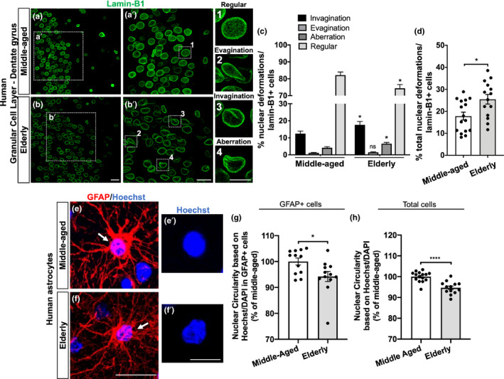
FIGURE 6.

Neural cells, including astrocytes, from the granular cell layer of the human hippocampus undergo nuclear abnormalities upon aging. (a, b) Distinct nuclear morphological profiles were evaluated, such as regular (1), evagination (2), invagination (3), and aberration (4) at the hippocampal granular cell layer in post‐mortem human tissue from middle‐aged and elderly donors based on lamin‐B1 staining. Scale bars, 20 µm in (b) and (b′); 10 µm in (4). (c) Elderly donors exhibited a higher incidence of invaginated (p = 0.0494) and aberrant nuclei (p = 0.0243), resulting in a decreased proportion of regular nuclei (p = 0.0102), compared with the middle‐aged group. n = 16 and 14 individuals for middle‐aged and elderly groups, respectively. (d) Elderly donors showed an increased proportion of total nuclear deformations (ie, evagination + invagination + aberration) (p = 0.0119). n = 16 and 14 individuals for middle‐aged and elderly groups, respectively. (e–g) Nuclear circularity was quantified based on Hoechst or DAPI staining in post‐mortem human tissue from middle‐aged (e–e′) and elderly donors (f–f′). Elderly donors presented a reduced nuclear circularity based on the number of GFAP+cells (g; p = 0.0243; n = 12 individuals for both age groups) and on the total number of cells (h; p < 0.0001; n = 15 and 13 individuals for middle‐aged and elderly groups, respectively). Scale bars, 20 µm in (f) and 10 µm in (f′). Significance was determined using unpaired t test with Welch's correction. Error bars represent ± SEM. Individual data points are plotted and represent individual donors
