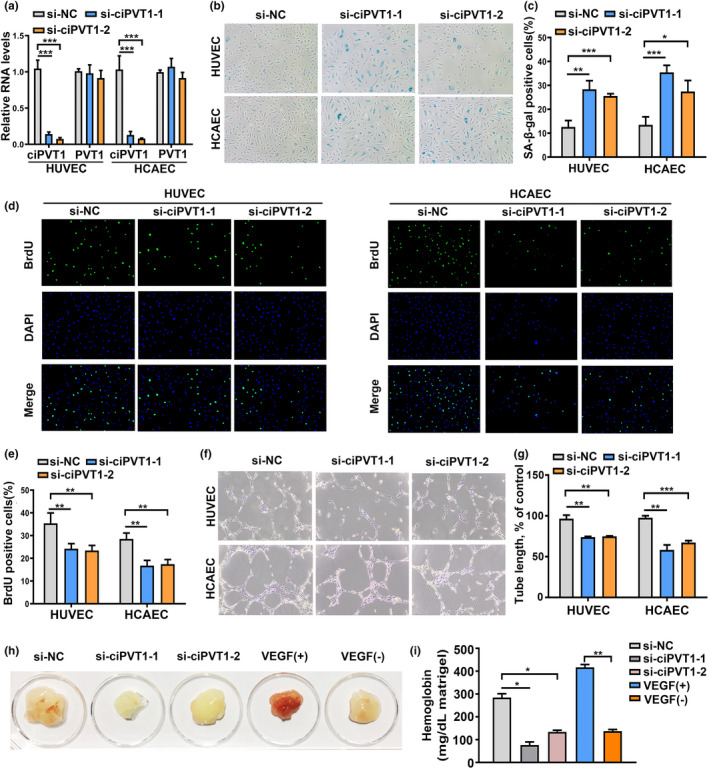
FIGURE 2.

Knockdown of ciPVT1 promoted senescence, inhibited proliferation, and angiogenesis of ECs. (a) RT‐qPCR analysis of ciPVT1 and PVT1 RNA expression in proliferating ECs 3 days after transfection with si‐NC, si‐ciPVT1‐1, and si‐ciPVT1‐2. (b) Representative photographs of SA‐β‐gal staining of ECs transfected with si‐NC, si‐ciPVT1‐1, and si‐ciPVT1‐2. (c) The SA‐β‐gal‐positive cells were counted and presented as percentage of total cells. (d) Representative images of the indicated cells stained with DAPI (blue fluorescence) and BrdU, as a measurement of DNA synthesis (green fluorescence) in si‐NC‐, si‐ciPVT1‐1‐, and si‐ciPVT1‐2‐transfected ECs. (e) The BrdU‐positive cells were counted and presented as percentage of total cells. (f and g) Representative micrographs and statistical summary of in vitro Matrigel assays in si‐NC‐, si‐ciPVT1‐1‐, and si‐ciPVT1‐2‐transfected ECs. (h) CiPVT1 and angiogenesis in vivo. Athymic nude mice received a subcutaneous injection of Matrigel plugs supplemented with saline, VEGF, or mixed with HUVECs transfected with si‐NC, si‐ciPVT1‐1, and si‐ciPVT1‐2. Representative gross appearance of Matrigel plugs. (i) Quantification of hemoglobin (Hb) in the homogenized Matrigel plugs. Data are presented as mean ± SD; *p < 0.05, **p < 0.01, ***p < 0.001
