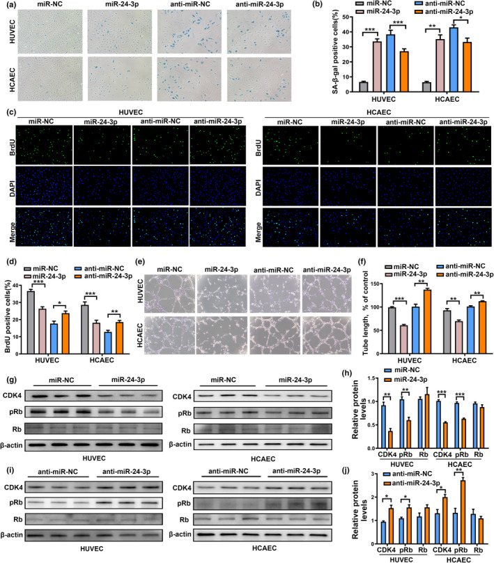
FIGURE 4.

MiR‐24‐3p promoted senescence, reduced proliferation, and impaired the capillary tube network formation ability of ECs. (a) Representative photographs of SA‐β‐gal staining of ECs transfected with miR‐24‐3p or miR‐NC, and anti‐miR‐24‐3p or anti‐miR‐NC. (b) The number of senescent cells was counted and presented as percentage of SA‐β‐gal‐positive cells. (c) Representative images of cells stained with DAPI and BrdU in miR‐24‐3p‐ or miR‐NC‐, and anti‐miR‐24‐3p‐ or anti‐miR‐NC‐transfected ECs. (d) The BrdU‐positive cells were counted and presented as percentage of total cells. (e and f) Representative micrographs and statistical summary of in vitro Matrigel assays in miR‐24‐3p‐ or miR‐NC‐, and anti‐miR‐24‐3p‐ or anti‐miR‐NC‐transfected ECs. (g and h) CDK4, pRb, Rb protein expression, and intensity ratio between CDK4, pRb, Rb, and β‐actin in ECs transfected with miR‐24‐3p or miR‐NC. (i and j) CDK4, pRb, Rb protein expression, and intensity ratio between CDK4, pRb, Rb, and β‐actin in ECs transfected with anti‐miR‐24‐3p or anti‐miR‐NC. Data are presented as mean ± SD; *p < 0.05, **p < 0.01, ***p < 0.001
