FIGURE 1.

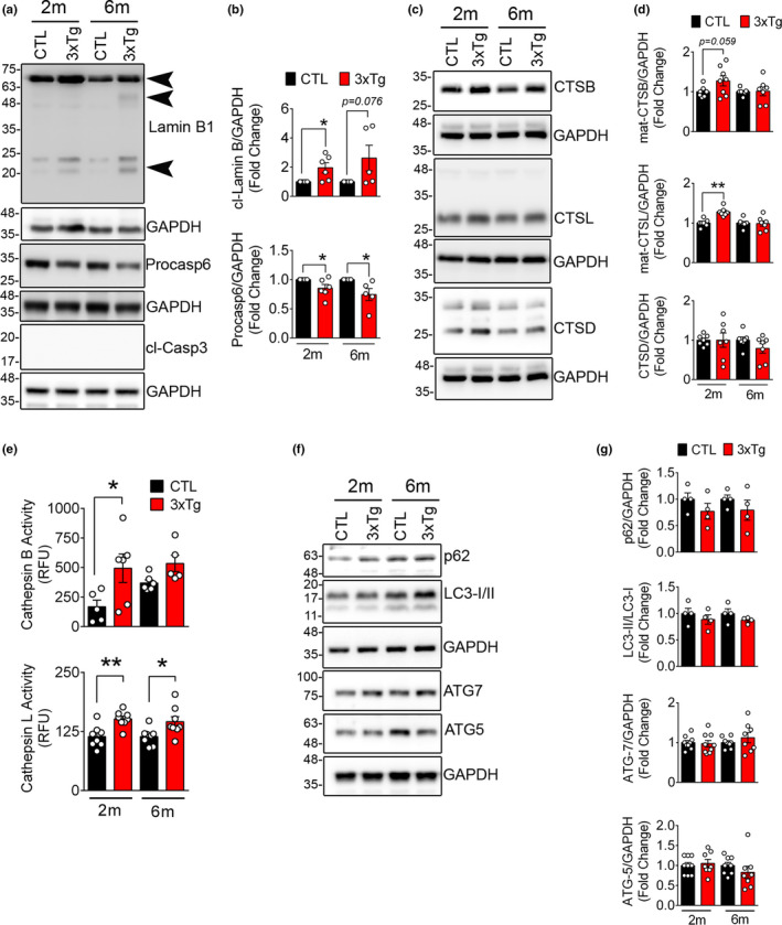
Lamin B1 degradation and upregulation of lysosomal cathepsins in 3xTg mouse hippocampus tissue (a) Representative Western blots showing the expression of lamin B1, pro‐casp6, and cl‐Casp3 in hippocampal lysates from CTL and age‐matched 3xTg mouse. (b) Quantification of cleaved lamin B1 (21 kDa) and pro‐casp6 is shown in 2 months (2m) and 6 months (6m) old mice. (n = 5–6 mice/group), data shown as ±SEM, *p < 0.05. Increased cleaved lamin B1 in 3xTg mice is detected by appearance of a 46 kDa and a 21 kDa (arrowhead). As reported previously, a significant decrease in pro‐casp6 protein was confirmed in 3xTg mouse. We did not detect any indication of apoptosis in these mice as assessed by lack of cl‐Casp3. (c) Representative Western blots showing lysosomal cathepsins in 3xTg mouse hippocampus were quantified using densitometry. (d) Bar graph shows increased levels of CTSB and CTSL in 3xTg mouse, although CTSD levels remained unchanged. (e) Enzymatic activity of CTSB and CTSL activity in CTL and age‐matched 3xTg hippocampal lysates. (f) Markers of autophagy progression were assessed using immunoblotting. (g) No significant change in autophagy progression was observed in this model
