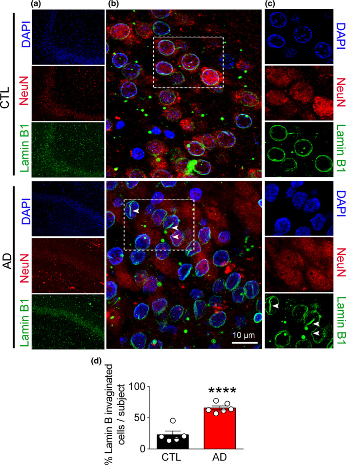
FIGURE 2.

Nuclear lamina damage is detected by invagination and focal loss of Lamin B1 in human AD brain. (a) Confocal microscopic micrographs depicting histological examination of hippocampal and medial temporal cortex from autopsy samples: LB1 (green), NeuN (red), and DAPI (blue). (b and c) Higher magnifications of the selected regions from A are shown (scale bar = 10 µm). (d) Quantification of damaged neuronal nuclei, as identified with their coffee‐bean nuclei immunolabeled with LB1 is shown. Cell counting was performed using ImageJ. A minimum of 109 cells were counted for each sample and the number of invaginated cells were expressed as % of total cells. (CTL = 5 and AD = 6 samples). Data reported as mean ±SEM. **** represents p < 0.0001
