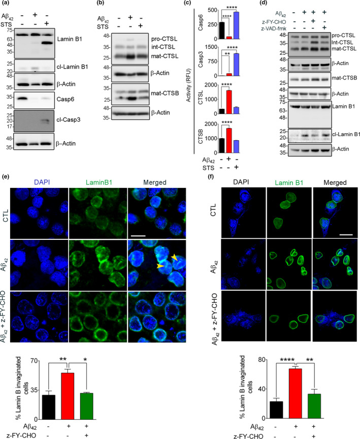
FIGURE 4.

Aβ42‐induced lamin B1 cleavage is independent of apoptosis. To differentiate the contribution of apoptosis and autophagy to lamin B1 cleavage, SH‐SY5Y cells were exposed to Aβ42 (5 µM, 16 h) and STS (0.5 µM, 6 h). (a) Western blots showing the appearance of a lamin B1 46 kDa fragment that was only observed in STS‐treated cells. This was associated with a significant loss of pro‐casp6 protein and presence of cleaved Casp3, indicating the involvement of caspases. The 46 kDa fragment of lamin B1 and pro‐casp6 were not observed in Aβ42‐treated cells and no cleaved Casp3 was detected in these cells. (b) Representative immunoblots examining the changes in lysosomal cathepsin protein level. (c) Enzymatic activity assays for Casp6, Casp3, CTSL, and CTSB indicated the induction of caspases in STS‐treated cells and preferential activation of cathepsins in Aβ42‐treated cells. **p < 0.01 and **** p < 0.0001. (one‐way ANOVA/Tukey's post hoc). (d) Western blot confirming the involvement of CTSL in lamin B1 damage in rat primary hippocampal neurons when exposed to Aβ42 (5 µM, 16 h). Overnight pre‐treatment of neurons with CTSL inhibitor alleviated lamin B1 damage, but a pan‐caspase inhibitor z‐VAD‐fmk (20 µM) did not have any effect. (e) Confocal micrographs depicting cultured rat hippocampal neurons labeled for lamin B1 status in Aβ42 toxicity. LB1 invagination was observed in this model but was prevented significantly by CTSL inhibitor. The bar graph represents the percentage of invaginated/nicked neuronal LB1. A minimum of 130 cells/condition were analyzed. (f) The sensitivity of neuronal LB1 damage and NL invagination was also confirmed in mouse primary cortical neurons. This was preventable by pre‐treatment with CTSL inhibitor. An average of 56 cells were examined/experimental group. Results are mean ±SEM, *p < 0.05, **p < 0.01 and ****p < 0.0001, one‐way ANOVA/Tukey's post hoc, scale bar = 10 µm
