
Figure 1.
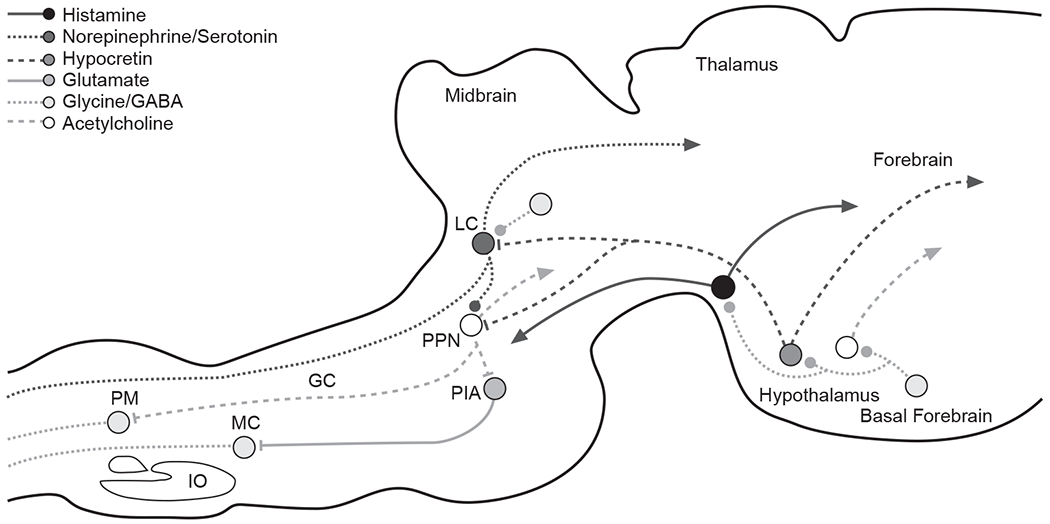
Synaptic Relationships Underlying the Loss of Consciousness and Correlated Loss of Muscle Tone in Normal Sleepa
aThis simplified drawing of a sagittal section of a cat’s brain shows some of the major connections that play an important role in sleep control. It also illustrates the complexity of the synaptic relationships underlying the loss of consciousness and correlated loss of muscle tone that characterize normal sleep. Systemic drug application can affect sleep through actions on any one of these synapses as well as on many other synapses not illustrated. Lines ending in solid dots indicate inhibitory output. Lines ending in arrows indicate excitatory output.
Abbreviations: GABA = γ-aminobutyric acid, GC = nucleus gigantocellularis, IO = inferior olive, LC = locus ceruleus, MC = nucleus magnocellularis, PIA = pontine inhibitory area, PM = nucleus paramedianus, PPN = pedunculopontine nucleus.