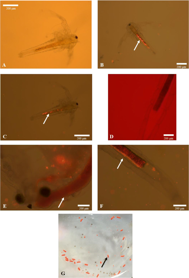
Figure 3.
Plastic fluorescent red polymer microspheres (FRM, 1–5 μm diameter) ingested, egested and adhere (white arrows) to Artemia franciscana nauplii and juveniles and their aggregation to faecal pellets (black arrow), visualized using fluorescence microscopy. Digestive tract of nauplii after 2 days of exposure to different FRM concentrations: A CTRL (0 mg L−1), B FRM0.4 (0.4 mg L–1) and C FRM1.6 (1.6 mg L−1). Digestive tract of juvenile after 5 days of exposure to different FRM concentrations: D CTRL (0 mg L−1), E FRM0.4 (0.4 mg L−1) and F FRM1.6 (1.6 mg L−1). FRM aggregation into faecal pellets and sunk at the bottom of glass flaks (G).

