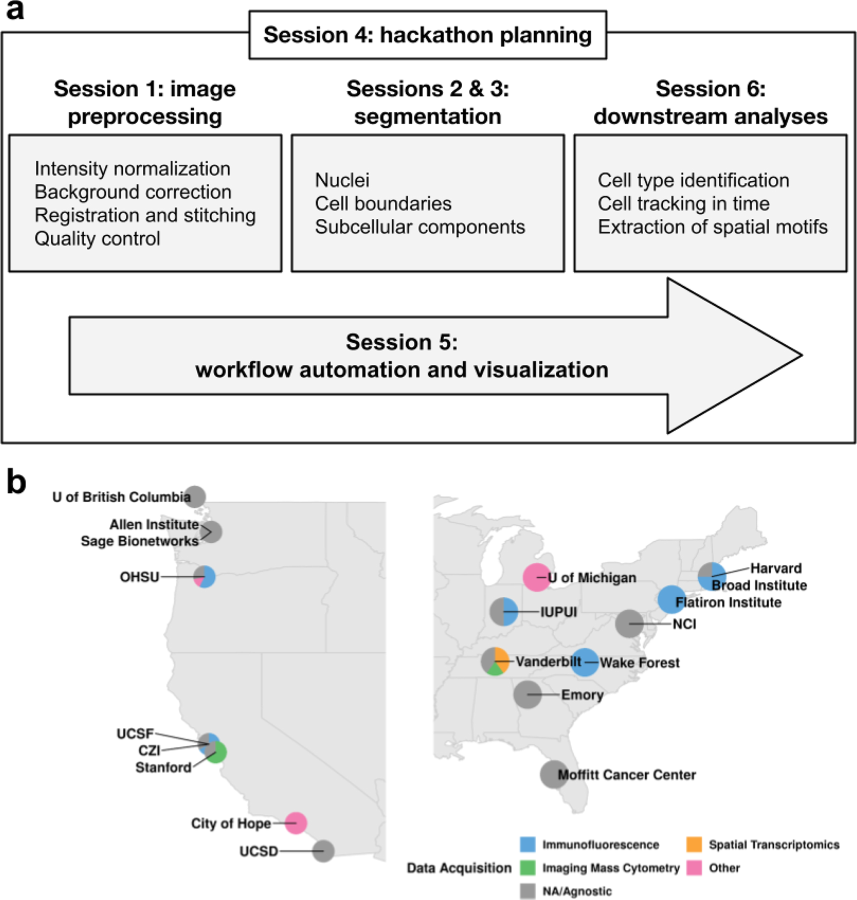
Figure 1: Image analysis workshop structure and participation.

a. The workshop agenda followed the steps of a canonical image processing workflow. Each box highlights the most prominent topics covered during each session. b. Institutes and data acquisition technologies represented by the workshop participants. Technologies marked “other” encompass electron microscopy and radiology, while “NA/Agnostic” refers to computational labs that don’t generate data.
