Figure 3.
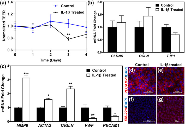
Long-term IL-1β treatment induces EndoMT in iBMECs. (a) TEER measurements indicate a drop in iBMEC TEER after 3 days of IL-1β treatment. **p < 0.01 compared to control at day 3 (unpaired t test). (b) Gene expression levels of tight junction proteins are not altered after 3 days of treatment with IL-1β (unpaired t test). (c) Endothelial markers (VWF and PECAM1) are downregulated, whereas mesenchymal markers (ACTA2 and TAGLN) and MMP9 are upregulated as a result of IL-1β treatment for 3 days. *p < 0.05, **p < 0.01, and ***p < 0.001 (unpaired t test). For (b, c), fold change is relative to non-treated control. Error bars represent standard deviation in (a–c). (d–g) Immunocytochemistry of iBMECs treated with IL-1β for 3 days shows downregulation of PECAM-1 in treated iBMECs (e) compared to control samples (d), while SM-22α expression is higher in IL-1β treated iBMECs (g) compared to control cells (f). Images are maximum intensity projections of confocal z-stacks, and scale bars indicate 50 μm.

