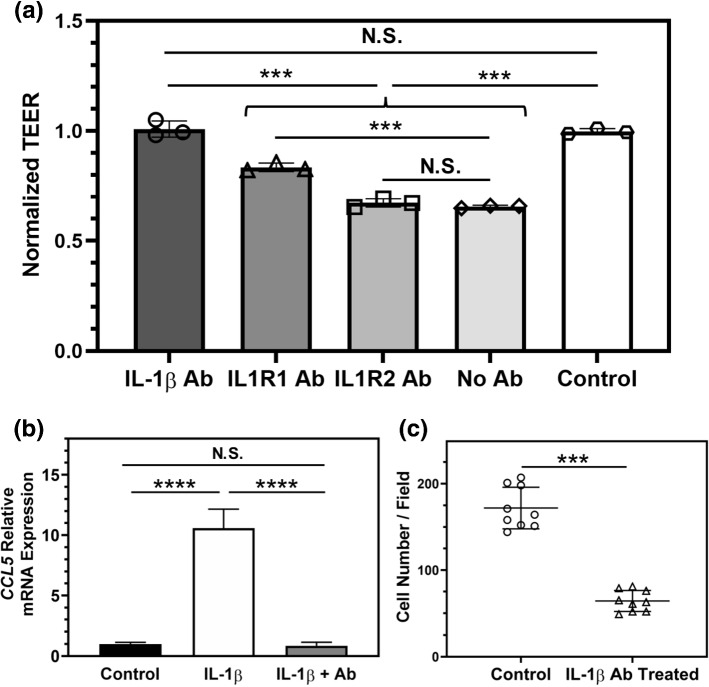
Figure 6.
Neutralizing IL-1β reduces barrier disruption and transmigration of 231BR cells. (a) TEER measurements indicate that blocking IL-1β with a monoclonal antibody (IL-1β Ab) is most effective at preventing iBMEC barrier disruption as a result of IL-1β addition, as compared to blocking IL-1β receptors on the iBMECs using IL1R1 or IL1R2 blocking antibodies. ***p < 0.001 (ordinary one-way ANOVA and Tukey's multiple comparisons test). Control samples received no IL-1β. Each data point shows the normalized TEER of a technical replicate. Results are representative of two independent experiments. (b) Addition of the IL-1β monoclonal antibody to the apical chamber of cell culture inserts prevents activation of astrocytes on the basolateral side after apical IL-1β treatment of iBMECs for 24 h. CCL5 gene expression was used as a measure of astrocyte activation. ****p < 0.0001 (ordinary one-way ANOVA and Tukey's multiple comparisons test). Three technical replicates were used to generate the plot. Results are representative of two independent experiments. (c) Incubating 231BR cells in the BBB-on-a-chip devices with astrocytes in the presence of IL-1β monoclonal antibody in the apical channel of the devices significantly reduces the number of cancer cells that transmigrate across the iBMECs, compared to the control group that did not receive IL-1β monoclonal antibody. ***p < 0.001 (unpaired t test). Each data point represents the number of transmigrated 231BR cells across the iBMECs in a confocal image. Three images per device and three devices per condition were used to generate the plot. Error bars represent standard deviation in (a–c).

