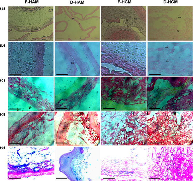
Figure 2.
Histological analysis of H&E stained sections of fresh and decellularized membranes reveals complete removal of epithelial cell layer and trophoblast cell layer in D-HAM and D-HCM after decellularization process. (a) ×10 magnification, scale bar 100 µm. (b) ×40 magnification, scale bar 50 µm. In contrast, it is visible from the stained sections that F-HAM and F-HCM containing a well distinguished epithelial and trophoblast cell layers. After the decellularization process, D-HAM and D-HCM preserve the ECM, and a complete absence of nuclei can be seen in both D-HAM and D-HCM. (c) Sirius red staining showed no signficant change in the distribution and quantity of collagen after decellularization. (d) Masson trichrome staining showing intact appearance of collagen and elastin with no obvious disruptions to collagen and elastin after decellularization. (e) Alcian Blue pH-1 staining demonstrated conserved GAGs after decellularization as compared to fresh membranes. ECL, Epithelial Cell Layer; BM, Basement Membrane; S, Stroma; TCL, Trophoblast Layer; RL, Reticular Layer.

