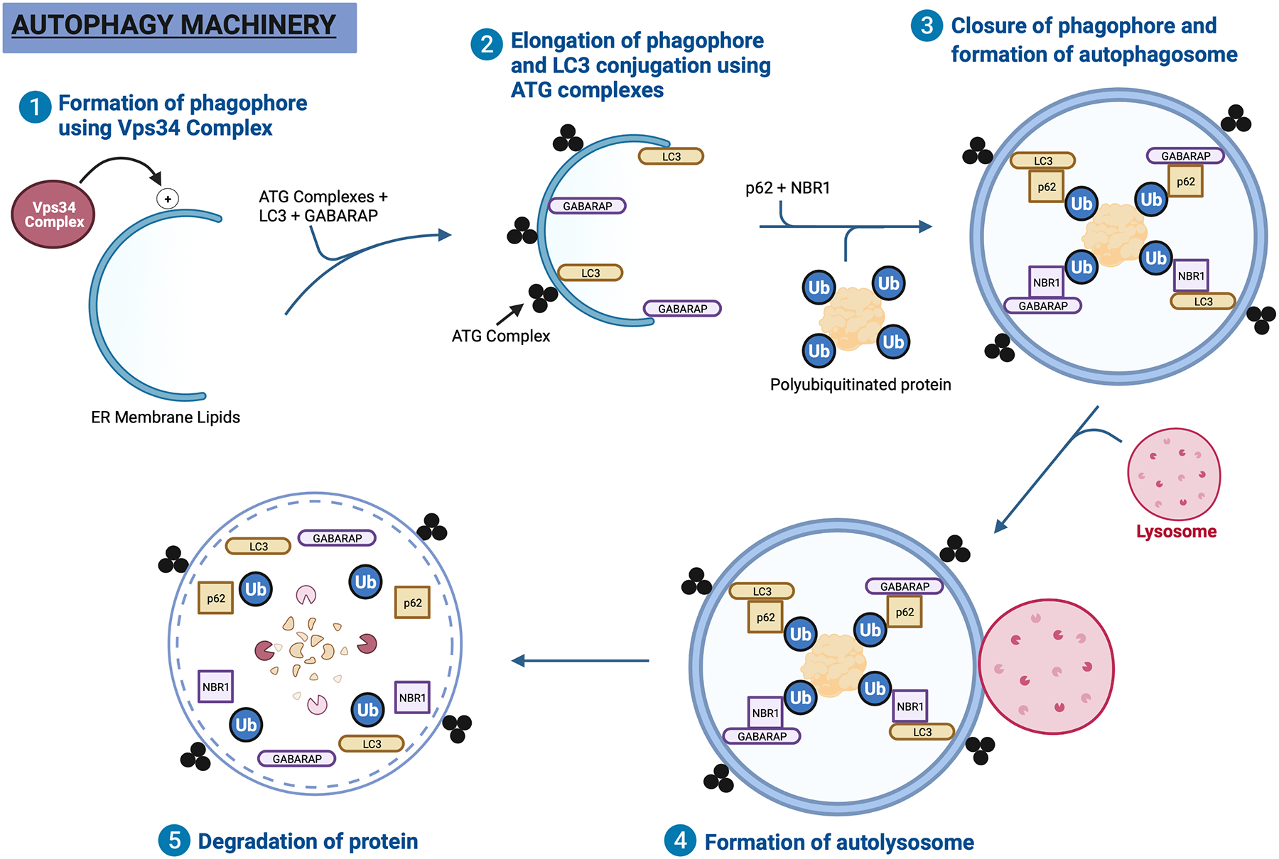
Figure 2. Autophagy machinery.

Step 1, the phagophore is synthesized from ER membrane lipids upon activation by the Vps34 Complex. Step 2, the phagophore is elongated with the help of structural proteins such as LC3 and GABARAP. These Atg8 family of proteins are attached to the phagophore with the help of ATG complexes. Step 3, p62 and NBR1 serve as cargo receptor proteins that carry polyubiquitinated proteins destined for degradation to the phagophore. Once the phagophore closes itself around the protein, it is known as an autophagosome. Step 4, a neighboring lysosome then fuses with the autophagosome to create and autolysosome. Step 5, the polyubiquitinated protein is degraded using lysosomal enzymes.
