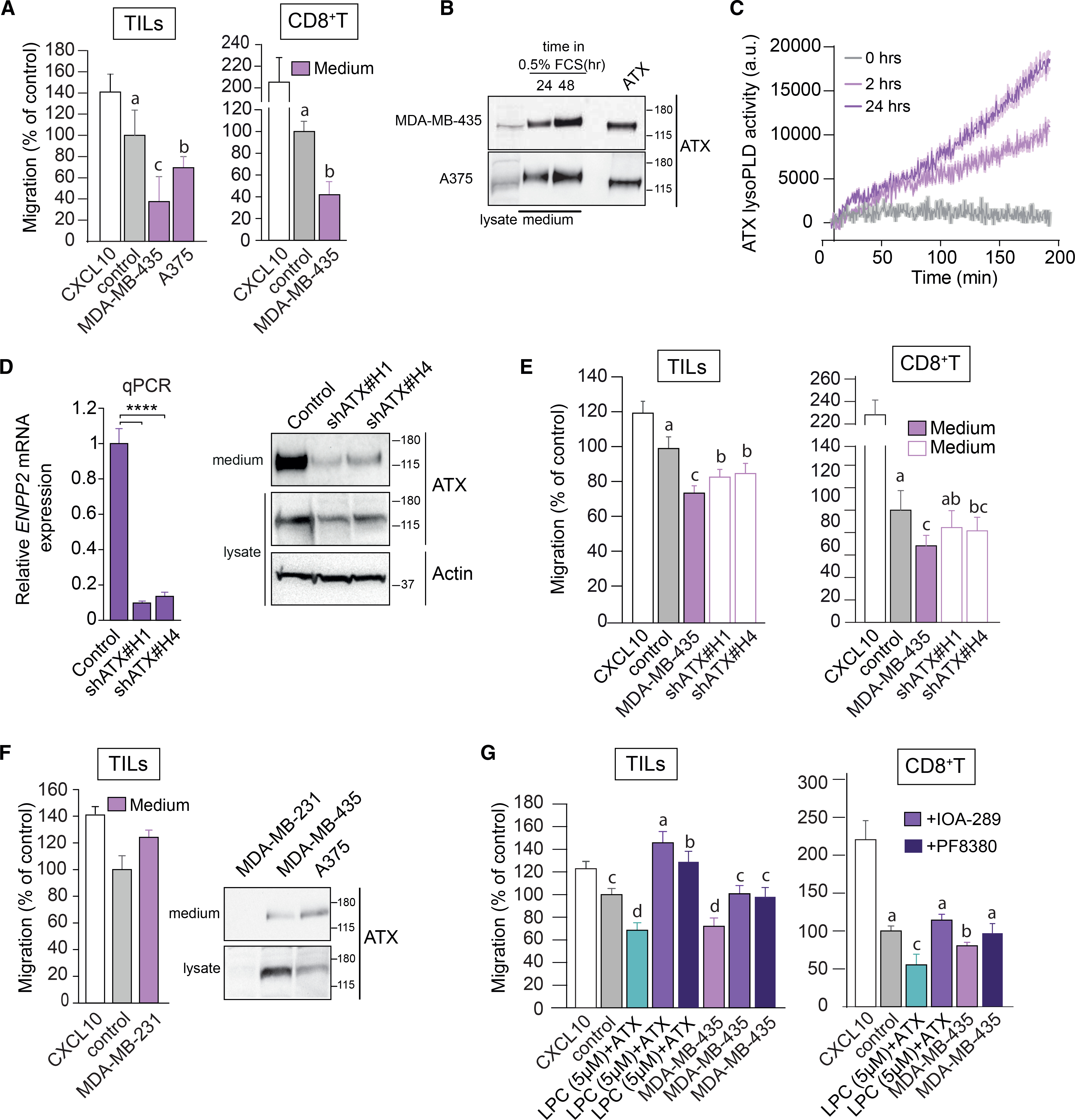
Figure 2. ATX secreted by melanoma cells repels TILs and peripheral CD8+ T cells.

(A) Melanoma-conditioned medium from MDA-MB-435 and A375 cells (collected after 24 h) is chemorepulsive for TILs and blood-derived CD8+ T cells. Experimental conditions as in Figure 1.
(B) Immunoblot showing ATX expression in medium and cell lysates of MDA-MB-435 and A375 melanoma cells. Cells were incubated in DMEM with 0.5% FCS for 24 or 48 h. Recombinant ATX (20 nM) was used as positive control (right lane).
(C) LysoPLD activity accumulating in melanoma-conditioned media over time. Medium from MDA-MB-435 cells was collected after 2 and 24 h, and lysoPLD activity was measured as choline release from added LPC(18:1).
(D) ATX (ENPP2) mRNA expression (relative to cyclophilin) in control and ENPP2-depleted MDA-MB-435 cells stably expressing short hairpin RNAs (shRNAs) against ATX. Maximal ENPP2 knockdown was obtained with shRNA 1 and 4 (of 5 different hairpins). Data represent the mean ± SEM of three independent experiments using triplicate samples; ****p < 0.0001 (unpaired Student’s t test). Right: immunoblot analysis of ATX expression using shRNA 1 and 4. Actin was used as loading control.
(E) Melanoma-conditioned medium from ATX knockdown MDA-MD-435 cells (collected after 24 h) lacks chemorepulsive activity for CD8+ T cells and TILs.
(F) Conditioned media from ATX-deficient MDA-MB-231 breast carcinoma cells lack chemo-repulsive activity for TILs compared to media from ATX-expressing melanoma cells (MDA-MB-435 and A375; cf. A). Right panel: ATX immunoblots from the indicated media and cell lysates.
(G) ATX inhibition restores the migration TILs and CD8+ T cells exposed to melanoma cell-conditioned media. Cells were plated at day 0 in medium containing 10% FCS. After 16 h, cells were exposed to medium containing 0.5% FCS and ATX inhibitors (PF-8380 or IOA-289). Conditioned media were collected after 24 h.
(A and D–G) Representative data of three independent experiments each performed in triplicate. Values are expressed as mean ± SEM; bars annotated with different letters were significantly different according to Fisher’s least significant difference (LSD) test (p ≤ 0.05) after ANOVA.
