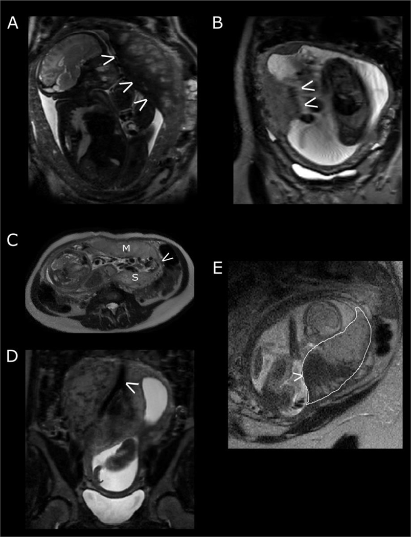
Figure 3:
Physiological factors affecting T2-weighted imaging. (A) fetal compression against the placenta resulting in a reduction in signal intensity. (B) artefact exacerbated by maternal respiratory motion. The fetal side of the placenta does not appear to have a distinct border. (C) Placenta with succenturiate lobe. M represents the main placental disc, S the smaller succenturiate lobe and the arrow pointing to vessels joining the two together. (D) the maternal aorta bifurcating into the right and left common iliac arteries. (E) The effect of a contraction on placental signal intensity. The placenta is outlined in white. Note the large low signal intensity area (arrow) and that the peripheral placenta is thicker than expected with less tapering at the edges.

