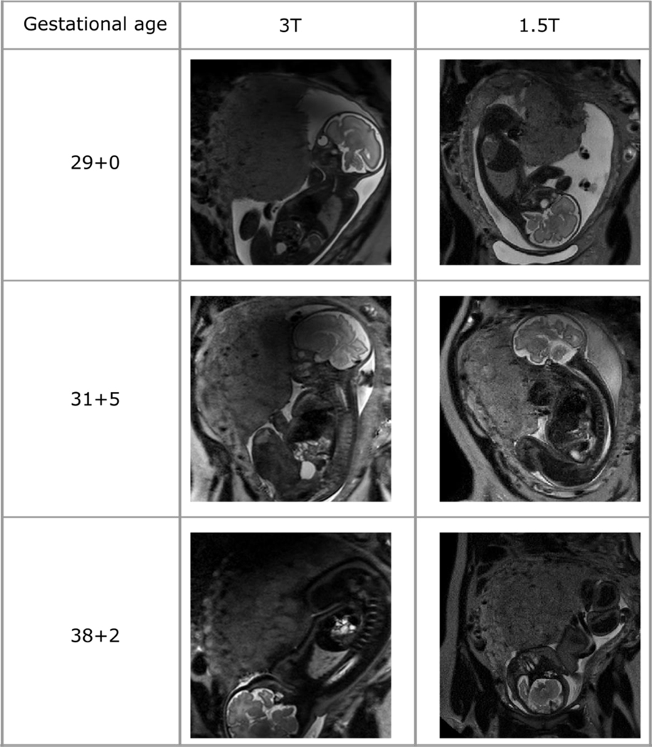
Figure 4:
T2-weighted placental imaging in maternal coronal plane performed on the same day at two different field strengths (3T and 1.5T) for three women. A disadvantage of imaging at 3T is noted where some diffuse low signal overlying the superior aspects of the placenta can be seen, secondary to magnetic field inhomogeneity.

