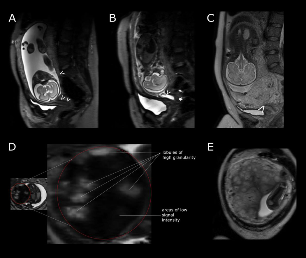
Figure 5:
T2-weighted imaging in pregnancy complications. (A) maternal sagittal plane illustrating a normal cervical length. A single arrow at the leading edge of the placenta and double arrows demonstrating tubular structure of the cervix of normal cervical length. (B) Arrow at the internal cervical os (demonstrating cervical widening or funnelling) and a short section of closed cervix (star) in a woman at risk of preterm delivery at 23 weeks’ gestation. (C) Placenta praevia completely covering the internal cervical os (white arrow). (D) Placenta in a pregnancy complicated by preeclampsia at 28 weeks’ gestation. (E) Placenta in a pregnancy complicated by chronic hypertension at 26 weeks’ gestation.

