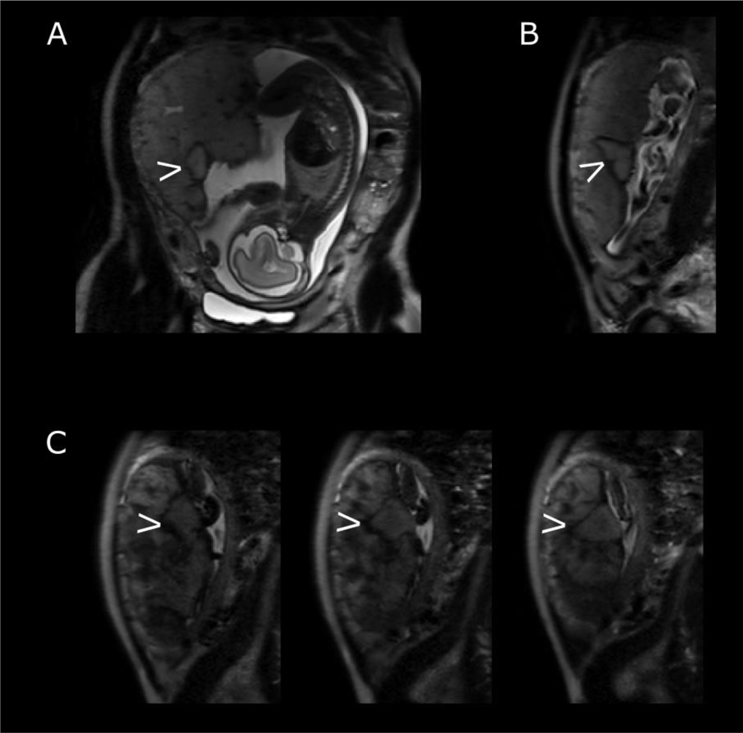
Figure 6:
Features on T2-weighted imaging confirmed by histological examination. Maternal coronal (A) and sagittal (B) planes with arrow indicating well circumscribed focal lesion with low signal periphery at 26 weeks’ gestation. This was confirmed as an evolving haematoma on histological examination after delivery. (C) Series of T2-weighted imaging in maternal sagittal plane at 30 weeks’ gestation with arrow indicating well circumscribed area below umbilical cord insertion. Histology revealed this to be an infarct.

