Figure 3.
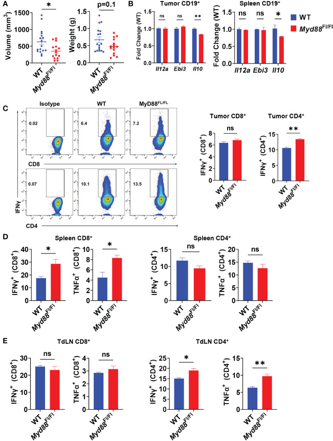
PDAC influences B cell IL-35 expression in vivo in a MyD88-independent manner. (A) Measured volumes and weights of orthotopic KPC4662 tumors from Cd19 +/+; Myd88 Fl/Fl (WT) and Cd19 Cre/+; Myd88 Fl/Fl (Myd88 Fl/Fl) mice at day 21 post-tumor injection. N=17. Cumulative of 3 individual experiments. (B) RT-PCR analysis of Il12a, Ebi3, and Il10 expression from CD19+ B cells derived from tumors and spleens of Cd19 +/+; Myd88 Fl/Fl and Cd19 Cre/+; Myd88 Fl/Fl mice at day 21 post-tumor injection. N=5. (C) Intracellular flow cytometry of IFNγ and TNFα in CD8+ and CD4+ T cells derived from tumors of Cd19 +/+; Myd88 Fl/Fl (WT) and Cd19 Cre/+; Myd88 Fl/Fl (MyD88Fl/Fl) KPC4662 orthotopic tumor-bearing mice at day 21 post-tumor injection. N=4. (D) Intracellular flow cytometry of IFNγ and TNFα in CD4+ and CD8+ T cells derived from spleens of Cd19 +/+; Myd88 Fl/Fl and Cd19 Cre/+; Myd88 Fl/Fl KPC4662 orthotopic tumor-bearing mice at day 21 post-tumor injection. N=4. (E) Intracellular flow cytometry of IFNγ and TNFα in CD4+ and CD8+ T cells derived from spleens of Cd19 +/+; Myd88 Fl/Fl and Cd19 Cre/+; Myd88 Fl/Fl KPC4662 orthotopic tumor-bearing mice at day 21 post-tumor injection. N=4. Error bars indicate SEM; p values in (A–D) were calculated using t-test; p values in (B) were calculated using two-way ANOVA. NS, non-significant, *p<0.05, **p<0.005, ***p<0.001 Experiments were performed using 7–8-week-old mice of indicated genotypes.

