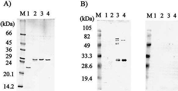
FIG. 2.
(A) Analysis by SDS-PAGE of purified recombinant proteins stained with Coomassie blue. (B) Immunoblot analysis using a pooled serum from NCC patients (left panel) or AE patients (right panel). Molecular size markers are indicated on the left. Lane 1, TRX; lane 2, rAg1; lane 3, rAg1V1; lane 4, rAg2.

