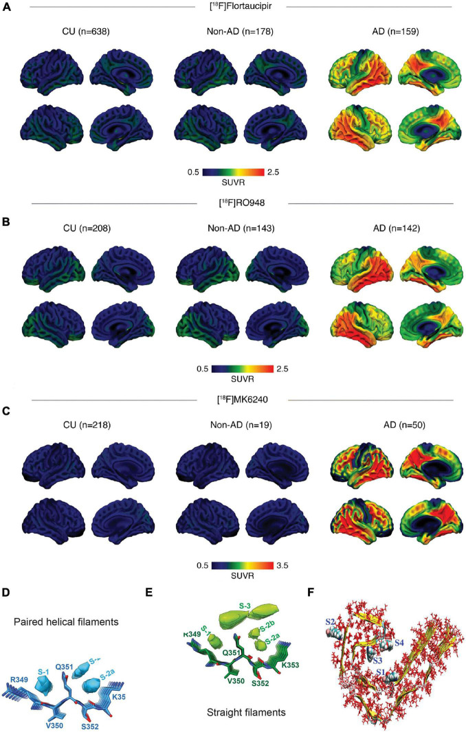
FIGURE 1.
In vivo and postmortem comparison of tau imaging probes in the human brain (A–C) multicenter comparison of positron emission tomography (PET) imaging using [18F]flortaucipir, [18F]RO948, and [18F]MK6240, standardized uptake value ratios (SUVRs) across all participants within diagnostic groups; non-demented controls (CU), Alzheimer’s disease (AD); reproduced from Leuzy et al. (2021) with permission from Springer Nature; (D,E) binding of APN-1607 (PM-PBB3) to tau-paired helical filaments and straight filaments is based on cryo-EM, top views and side views of the extra densities in the PM-PBB3 binding sites of paired helical filaments (D) and straight filaments (E) maps. The models of PM-PBB3 are shown near these extra densities at the same scale. Reproduced from Shi et al. (2021a) with permission from Springer Nature; and (F) various high-affinity binding sites of tau protofibril. The sites 1, 3, and 4 are termed core sites as they are buried inside the fibril, whereas site 2 is termed a surface site as it is exposed to a greater amount of solvent molecules. Reproduced from Murugan et al. (2018) with permission from American Chemical Society.

