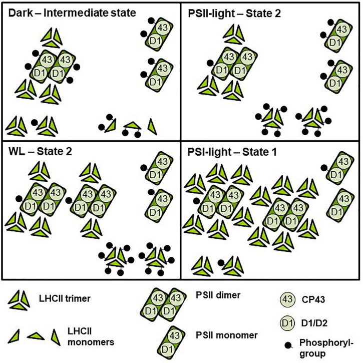
FIGURE 5.
Model depicting effects of light-quality or dark-white light shifts on assembly and phosphorylation states of thylakoid membrane proteins. The simplified scheme integrates the central biochemical data from Figures 2–4 and Supplementary Table S1. Symbol representations are defined at the bottom of the scheme. The number of protein complexes and of phosphoryl-groups per protein complex indicates relative accumulation and degree of phosphorylation between the four different conditions. Dark: Low accumulation of PSII super-complexes, moderate phosphorylation state of PSII-bound LHCII and free LCHII trimers, PSII monomers with strongly phosphorylated CP43. PSII-light – State 2: Low accumulation of PSII super-complexes, PSII monomers with strongly phosphorylated CP43, high phosphorylation state of LHCII trimers. WL: High accumulation of PSII super-complexes (mostly C2S1 and C2S2), PSII monomers with low to moderately phosphorylated CP43, very high phosphorylation of LHCII trimers. PSI-light – State 1: Very high accumulation of PSII super-complexes (including C2S2M2 to C2S1), low phosphorylation state of PSII-bound and free LHCII trimers and of PSII dimers. PSII monomers with largely dephosphorylated CP43. For functional implications of the different assembly and phosphorylation states, see “Discussion” section.

