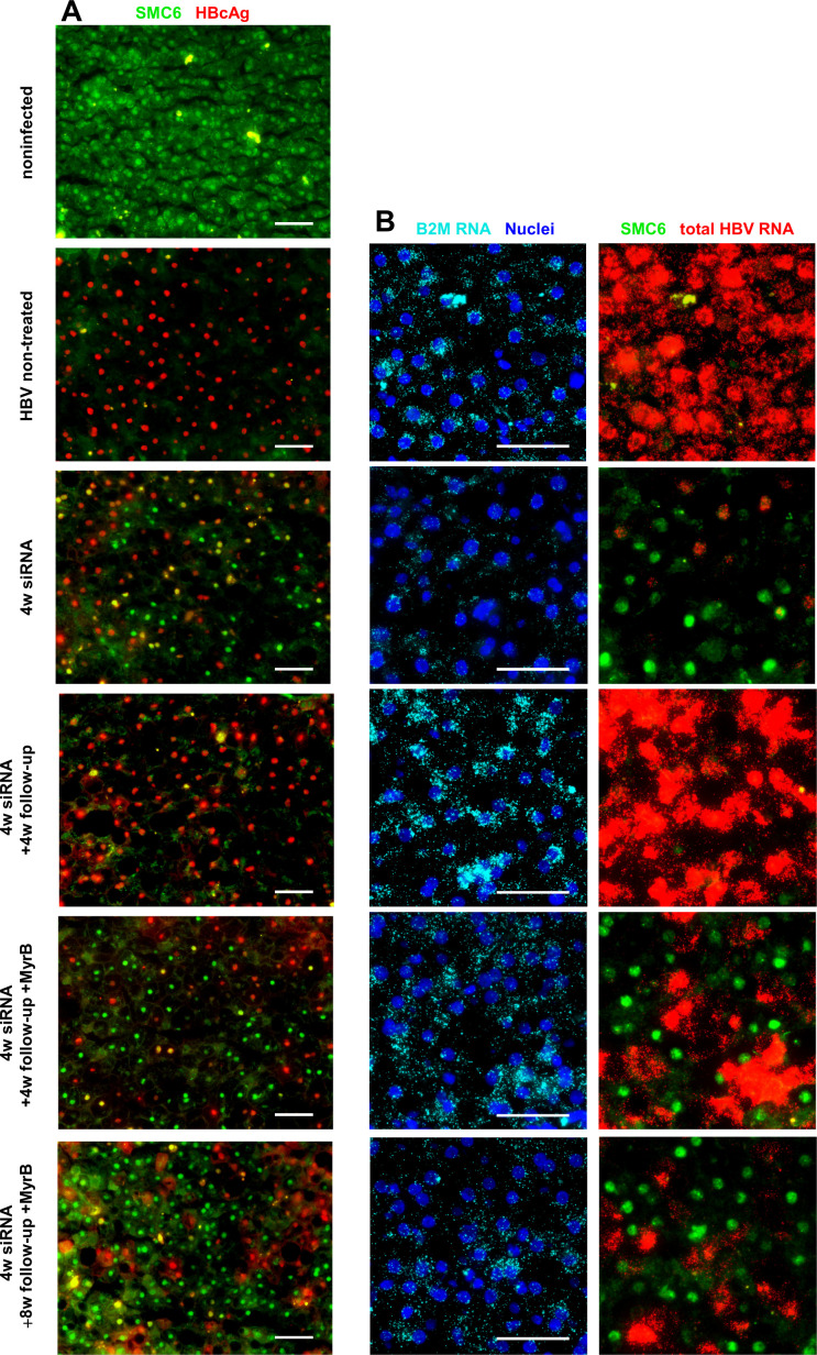
Figure 2.
siRNA treatment leads to the reappearance of SMC6 in hepatocytes negative for signs of active HBV replication. (A) Immunofluorescence costaining for HBcAg (red) and SMC6 (green) in cryopreserved liver sections. Shown are representative pictures of one mouse from every treatment group as indicated on the left-hand side. (B) Representative pictures of RNA in situ hybridisation for total HBV RNA combined with immunofluorescence staining for SMC6 protein. Merged pictures of nuclei stained with 4′,6-diamidino-2-phenylindole (blue) and B2M RNA as a marker for human hepatocytes (aqua) are shown in the left column; HBV RNA (red) and SMC6 (green) are shown in the right column. Scale bar 50 µm. MyrB, myrcludex-B; siRNA, small interferring RNA.

