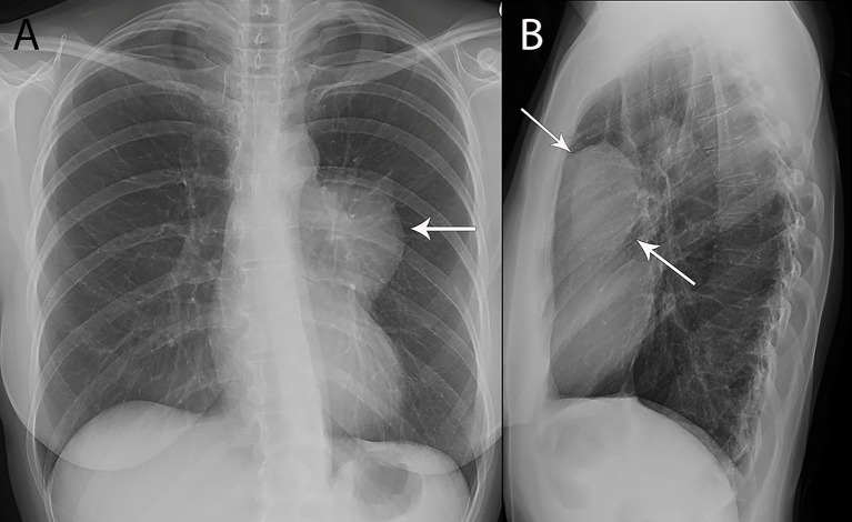
Figure 1.
53 year old woman with thymoma. (A, B) Frontal chest radiograph (A) shows left mediastinal contour abnormality (arrow) that results in loss of the silhouette of the upper left heart border. (B) The lesion is localized to the prevascular mediastinum in the retrosternal space (arrows) on the lateral chest radiograph (B).

