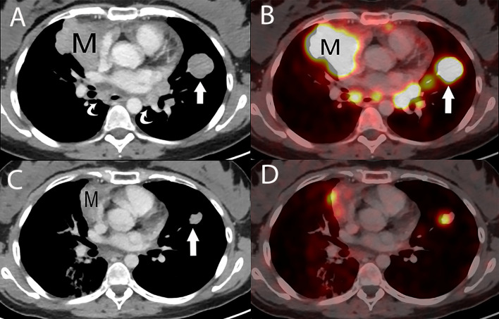
Figure 14.
29 year old woman with metastatic thymic carcinoma treated with peptide receptor radiotherapy (PRRT) with 177Lutetium. (A) Baseline contrast enhanced chest CT prior to PRRT treatment shows the primary prevascular mass (M), lung metastasis (straight arrow) and metastatic mediastinal lymphadenopathy (curved arrows). (B) Axial fused PET/CT scan at the same level as A, using a somatostatin analogue, Ga 68-DOTATATE, reveals DOTATATE uptake in the primary mass (M), lung metastasis (straight arrow) and mediastinal lymphadenopathy. (C) Contrast enhanced chest CT, following two peptide receptor radiotherapy (PRRT) with 177Lutetium sessions show an impressive partial response with a decrease in size of the primary mass (M) and of the lung metastasis (straight arrow), with resolution of the mediastinal lymphadenopathy. (D) Axial fused Ga 68-DOTATATE PET/CT scan at the same level as C, shows a corresponding marked decrease in DOTATATE uptake consistent with an impressive metabolic partial response.

