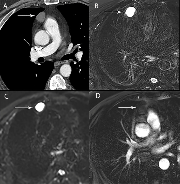
Figure 4.
67 year old man with right thymic cyst. (A) Contrast-enhanced CT shows a right prevascular mediastinal 2.2 cm lesion (arrow) with 34 Hounsfield units, which can represent solid or cystic lesion with proteinaceous material or hemorrhage. (B–D) MRI is useful to determine that this is a simple thymic cyst (arrow) with high signal intensity on T2 weighted (B) and short tau inversion recovery (STIR) (C) and no enhancement on the post contrast images (D).

