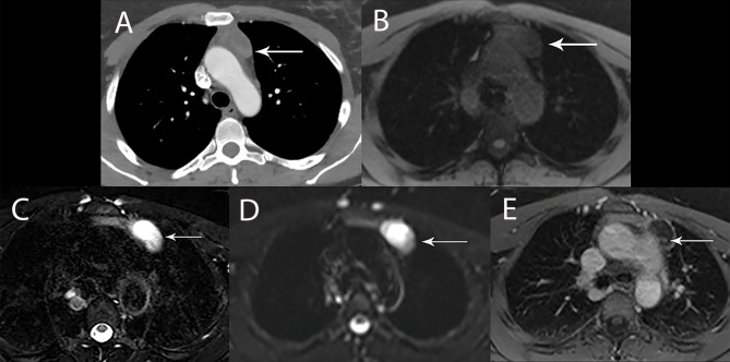
Figure 5.
44 year old woman with cystic thymoma. (A) Contrast enhanced CT shows solid component along the posterior aspect. MRI showed intermediate signal intensity on T1 weighted (B), high signal intensity on T2 weighted images (C) and DWI (D) consistent with cystic component. The small solid component (arrow) shows enhancement on post contrast T1 weighted images (E).

